358 posters, 97 topics, 122 sessions, 958 authors, 239 institutions
ePostersLive by SciGen Technologies S.A. All rights reserved.
79th PostGraduate Assembly in Anesthesiology 2025
December 12 - 15, 2025 | New York City

2212
The Enigma of Stanford Type I Aortic Dissection: Right Coronary Artery Involvement
Part of Topic
Cardiovascular - Critical Care
The Enigma of Stanford Type I Aortic Dissection:
Right Coronary Artery Involvement
Hannah Soliman, BA1;Adam Abbas, MD2; Amira Elfergani1; Nala Bughrara2; Sharan Prasad B.S.3; Omar Daghstani4; Farzana Afroze, MD2; Nibras Bughrara, MD2
1Albany Medical College, 2Albany Medical Center, Department of Anesthesiology, Jacobs School of Medicine and Biomedical Sciences3, Albany Medical Center, Department of Cardiology4
Introduction
This case highlights the complexity of managing extensive Type A aortic
dissection in patients with significant comorbidities. Although imaging did
not confirm coronary involvement, intraoperative right coronary artery
compromise required urgent grafting, further complicating the already
challenging surgical repair. Postoperative complete heart block, persistent
bleeding, and mixed shock states pointed toward evolving
cardiopulmonary compromise, and despite aggressive ICU management,
the patient progressed to cardiogenic shock and cardiac arrest.
Intra-arrest TEE demonstrated severe right heart failure, an
underrecognized complication following aortic surgery, underscoring the
importance of anticipating postoperative RV dysfunction in high-risk
patients with renal disease, major volume shifts, and myocardial strain.
Early recognition and consideration of mechanical circulatory support with
percutaneous coronary intervention in appropriate candidates may
improve outcomes, particularly in cases complicated by graft failure or
unusual coronary anatomy (5).
Visualization Discussion
49-year-old male with ESRD, HTN, HLD, OSA, and PUD presented with
acute tearing chest pain radiating to the back and right leg numbness.
Imaging revealed an Type A acute aortic dissection (TAAD) originating at
the aortic root measuring 4.7cm and ascending measuring 4.3 x 4.5cm.
Emergent surgical repair of the ascending aorta was performed; a
damaged RCA branch was noted after completion of the proximal
anastomosis requiring re-arrest and CABG. Preoperative and
postoperative EF were normal. CVICU course included bilateral pleural
effusions, pneumothorax, persistent bleeding near the pulmonary artery,
hypothermia, and leukocytosis. Vasopressor requirements were weaned
throughout the week, but remained on low dose dobutamine. A new RLL
opacity was found on X-ray. Bronchioalveolar lavage revealed numerous
mucous plugs. Intra-arrest TEE showed RCA graft failure and akinetic RV.
Resuscitative efforts were halted during CPR after discussion with the
family.
● Stanford type A aortic dissection (TAAD) is a cardiovascular emergency
with high morbidity and mortality requiring urgent surgical repair (1).
● Coronary artery involvement occurs in ~10-15% of cases and
significantly increases mortality (2,1).
● Right coronary artery (RCA) compromise is problematic, predisposing to
right ventricular dysfunction, arrhythmias, and cardiogenic shock (3,1).
● Ostial involvement may be missed on imaging, and intraoperative
findings may necessitate urgent RCA grafting (4,1).
● Early recognition and aggressive postoperative support are critical,
though outcomes remain poor in complex presentations (3,1).
Case Description
A
B
C
D
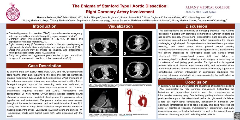
Figure 1 Axial CT Thorax Showing in Panel A from left to right the Brachiocephalic artery, Common Carotid artery, and Subclavian
Artery. The false lumen of the ascending aorta (Panel A), aortic arch (Panel B), and sinotubular junction (Panels C and D). The right coronary
artery is observed on the anterior surface of the heart (Panes C & D).
Figure 2 Transhepatic Lung View displaying fluid in the right
pleural sac, and fluid in the abdominal cavity. The thoracic spine
is visible at the most posterior segment of the image.
References:
In summary, this case underscores the challenges of managing extensive
TAAD complicated by right coronary involvement, highlighting the
limitations of preoperative imaging and the consequences of
intraoperative coronary injury. Despite timely grafting and comprehensive
postoperative support, the patient developed acute right ventricular failure,
a rare but highly lethal complication, particularly in individuals with
significant comorbidities such as renal disease. This case reinforces the
need for heightened vigilance, multidisciplinary coordination, and early
recognition of right ventricular dysfunction, as well as the potential role of
advanced circulatory support in select high-risk patients.
Conclusion
Intima
Media
Adventitia
Figure 3 An illustration of an aortic aneurysm.Components
taken from Biorender.com (6).