150 posters, 16 sessions, 294 authors, 70 institutions
ePostersLive by SciGen Technologies S.A. All rights reserved.
Obstetric Anaesthesia Annual Scientific Meeting 2024
Thursday 9 - Friday 10 May 2024, Birmingham

6289793
Mechanical thrombectomy of a pulmonary embolus in a pregnant patient with a severely impaired right ventricle
Poster Presenter
Authors
Part of Topic
Mechanical thrombectomy of a pulmonary embolus in a pregnant patient with a severely impaired right ventricle
J Whitehouse, J Hague, H El-Gendy, K Makam
Department of Anaesthesia, University Hospitals of Leicester NHS Trust, Leicester, UK
Introduction:
Pulmonary embolus (PE) has been shown to be the cause of 15% of maternal deaths1. Pregnant patients are at higher risk of PE due to venous stasis, the potential for vascular injury and an overall hypercoagulable state. Current treatment guidelines recommend thrombolysis with unfractionated heparin for patients with life-threatening maternal collapse. Thrombectomy is not mentioned in current guidance and given its increased prominence and reduced risk profile, may prove a viable alternative.
Whilst acute massive PE is treated with unfractionated heparin, thrombolysis is reserved for life threatening cases. Thrombolysis carries a 2-3% risk of bleeding and compared with studies on patients who had a mechanical thrombectomy the risk profile appears to favour thrombectomy2.
Thrombectomy demonstrates rapid and sustained recovery of echocardiography parameters2. Data comparing thrombectomy and thrombolysis outcomes in PE is sparce, however evidence from stroke patients suggests that in large vessel occlusion it can have improved outcomes3.
Case report:
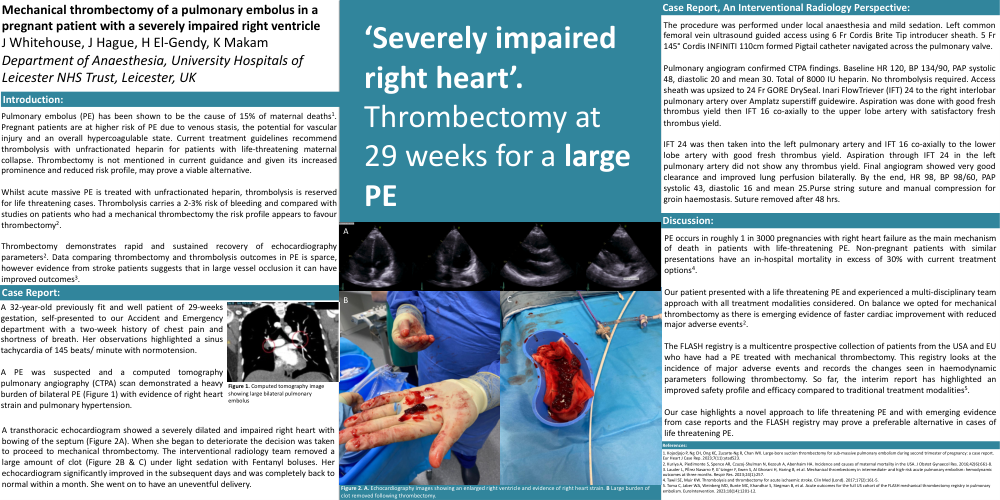
A 32-year-old previously fit and well patient of 29-weeks gestation, self-presented to our Accident and Emergency department with a two-week history of chest pain and shortness of breath. Her observations highlighted a sinus tachycardia of 145 beats/ minute with normotension.
A PE was suspected and a computed tomography pulmonary angiography (CTPA) scan demonstrated a heavy burden of bilateral PE (Figure 1) with evidence of right heart strain and pulmonary hypertension.
A transthoracic echocardiogram showed a severely dilated and impaired right heart with bowing of the septum (Figure 2A). When she began to deteriorate the decision was taken to proceed to mechanical thrombectomy. The interventional radiology team removed a large amount of clot (Figure 2B & C) under light sedation with Fentanyl boluses. Her echocardiogram significantly improved in the subsequent days and was completely back to normal within a month. She went on to have an uneventful delivery.
Case report, an interventional radiology perspective:
The procedure was performed under local anaesthesia and mild sedation. Left common femoral vein ultrasound guided access using 6 Fr Cordis Brite Tip introducer sheath. 5 Fr 145° Cordis INFINITI 110cm formed Pigtail catheter navigated across the pulmonary valve.
Pulmonary angiogram confirmed CTPA findings. Baseline HR 120, BP 134/90, PAP systolic 48, diastolic 20 and mean 30. Total of 8000 IU heparin. No thrombolysis required. Access sheath was upsized to 24 Fr GORE DrySeal. Inari FlowTriever (IFT) 24 to the right interlobar pulmonary artery over Amplatz superstiff guidewire. Aspiration was done with good fresh thrombus yield then IFT 16 co-axially to the upper lobe artery with satisfactory fresh thrombus yield.
IFT 24 was then taken into the left pulmonary artery and IFT 16 co-axially to the lower lobe artery with good fresh thrombus yield. Aspiration through IFT 24 in the left pulmonary artery did not show any thrombus yield. Final angiogram showed very good clearance and improved lung perfusion bilaterally. By the end, HR 98, BP 98/60, PAP systolic 43, diastolic 16 and mean 25.Purse string suture and manual compression for groin haemostasis. Suture removed after 48 hrs.
Discussion:
PE occurs in roughly 1 in 3000 pregnancies with right heart failure asthe main mechanism of death in patients with life-threatening PE. Non-pregnant patients with similar presentations have an in-hospital mortality in excess of 30% with current treatment options4.
Our patient presented with a life threatening PE and experienced a multi-disciplinary team approach with all treatment modalities considered. On balance we opted for mechanical thrombectomy as there is emerging evidence of faster cardiac improvement with reduced major adverse events2.
The FLASH registry is a multicentre prospective collection of patients from the USA and EU who have had a PE treated with mechanical thrombectomy. This registry looks at the incidence of major adverse events and records the changes seen in haemodynamic parameters following thrombectomy. So far, the interim report has highlighted an improved safety profile and efficacy compared to traditional treatment modalities5.
Our case highlights a novel approach to life threatening PE and with emerging evidence from case reports and the FLASH registry may prove a preferable alternative in cases of life threatening PE.
References:
1. Kojodjojo P, Ng CH, Ong KC, Zuzarte-Ng R, Chan WX. Large-bore suction thrombectomy for sub-massive pulmonary embolism during second trimester of pregnancy: a case report. Eur Heart J Case Rep. 2023;7(11):ytad523.
2. Kuriya A, Piedimonte S, Spence AR, Czuzoj-Shulman N, Kezouh A, Abenhaim HA. Incidence and causes of maternal mortality in the USA. J ObstetGynaecol Res. 2016;42(6):661-8.
3. Lauder L, PÈrez Navarro P, Gˆtzinger F, Ewen S, Al Ghorani H, Haring B, et al. Mechanical thrombectomy in intermediate- and high-risk acute pulmonary embolism: hemodynamic outcomes at three months. Respir Res. 2023;24(1):257.
4. Tawil SE, Muir KW. Thrombolysis and thrombectomy for acute ischaemic stroke. Clin Med (Lond). 2017;17(2):161-5.
5. Toma C, Jaber WA, Weinberg MD, Bunte MC, Khandhar S, Stegman B, et al. Acute outcomes for the full US cohort of the FLASH mechanical thrombectomy registry in pulmonary embolism. EuroIntervention. 2023;18(14):1201-12.