150 posters, 16 sessions, 294 authors, 70 institutions
ePostersLive by SciGen Technologies S.A. All rights reserved.
Obstetric Anaesthesia Annual Scientific Meeting 2024
Thursday 9 - Friday 10 May 2024, Birmingham

6301375
Emergency thoracotomy and caesarian section at 29 weeks gestation for suspected oesophageal rupture
Poster Presenter
Authors
Affiliations
Part of Topic
Emergency Thoracotomy and Caesarian Section at 29 Weeks Gestation for Suspected Oesophageal Rupture
Gardiner, M; Fletcher, J; Nishantha, T
Wrexham Maelor Hospital, Wales
INTRODUCTION
Thoracic complications involving the gastrointestinal (GI) tract are rare in pregnancy, with a literature search identifying limited case studies associated either with hyperemesis gravidarum during the first trimester (1), complications of congenital diaphragmatic hernias (2), or spontaneous pneumomediastinum.
CASE REPORT
Here, we present a case of suspected oesophageal rupture at 29 weeks gestation, treated with surgical management. A 44 year old gravida 3, para 1, 29/40, was admitted following repeat attendances, with a 2 week history of epigastric pain and worsening shortness of breath, on a background of previous Nissen’s fundoplication for gastro-oesophageal reflux disease 4 years prior.
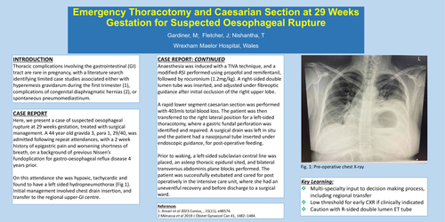
On this attendance she was hypoxic, tachycardic and found to have a left sided hydropneumothorax (Fig 1). Initial management involved chest drain insertion, and transfer to the regional upper-GI centre.
CASE REPORT: CONTINUED
Anaesthesia was induced with a TIVA technique, and a modified-RSI performed using propofol and remifentanil, followed by rocuronium (1.2mg/kg). A right-sided double lumen tube was inserted, and adjusted under fibreoptic guidance after initial occlusion of the right upper lobe.
A rapid lower segment caesarian section was performed with 403mls total blood loss. The patient was then transferred to the right lateral position for a left-sided thoracotomy, where a gastric fundal perforation was identified and repaired. A surgical drain was left in situ and the patient had a nasojejunal tube inserted under endoscopic guidance, for post-operative feeding.
Prior to waking, a left-sided subclavian central line was placed, an asleep thoracic epidural sited, and bilateral transversus abdominis plane blocks performed. The patient was successfully extubated and cared for post operatively in the intensive care unit, where she had an uneventful recovery and before discharge to a surgical ward.
References
1: Ansari et al 2023 Cureus, , 15(11), e48574.
2:Ménassa et al 2019 J Obstet Gynaecol Can 41, 1482–1484.
Fig. 1: Pre-operative chest X-ray
Key Learning: