191 posters, 5 topics, 20 sessions, 393 authors, 116 institutions
ePostersLive by SciGen Technologies S.A. All rights reserved.
Obstetric Anaesthesia Annual Scientific Meeting 2023
18 - 19 May, 2023 | EICC Edinburgh

5809678
Massive haemoptysis of unclear cause during pregnancy
Poster Presenter
Authors
Daniel George, James Matthews, Matteo De Martino, Mohamed Ragab, Ora Jesner, Emamoke Oteri
Affiliations
Part of Topic
Case report
Massive haemoptysis of unclear cause during pregnancy
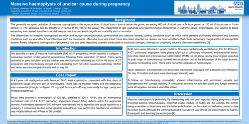
The generally accepted definition of massive haemoptysis is the expectoration of blood from a source below the glottis, exceeding 600 ml of blood over a 24 hour period or 150 ml of blood over a 1 hour period [1]. The sequelae can be thought of in terms of the risk to the airway, the impairment of gas exchange and haemodynamic compromise in extreme cases. Theoretically, any volume of blood exceeding that cleared from the bronchial tree per unit time can lead to significant morbidity and/ or mortality.
The differentials for massive haemoptysis are wide and include tracheobronchial, parenchymal and vascular lesions, cardiac conditions such as mitral valve disease, pulmonary embolism and systemic conditions such as vasculitis. Local infections such as pneumonia, often due to a viral insult, have also been reported as causes, as have conditions that cause secondary coagulopathy at telengectatic lesions. Rarely, idiopathic haemoptysis of pregnancy has also been described, possibly attributable to hormonal changes, whereby no underlying cause is ultimately established [2].
We describe a case of massive haemoptysis (750 ml) in pregnancy, which required a category 1 caesarean section under general anaesthesia following frank aspiration of blood. Twins were delivered in good condition and the mother was mechanically ventilated on ICU for 48 hours. A CT angiogram and bronchoscopy did not show a bleeding point nor other causative pathology. Mother and twins were discharged clinically well on day 10.
A 41 year old multigravida with twins of 36+3 weeks gestation, presented with five days of productive cough and one day of haemopytsis. There was no medical history and the pregnancy was uneventful (though on Aspirin 75 mg and Enoxaparin 40 mg antenatally for age, parity and multiple pregnancy).
Blood tests showed a haemoglobin of 120 g/L, platelets of 230 x 10^9/L and an international normalised ratio of 0.9. A CT pulmonary angiogram showed filling defects within the segmental bronchi. A witnessed episode of 500 ml frank haemoptysis led to aspiration and acute hypoxia so a category 1 caesarean section under general anaesthesia was performed. Mechanical ventilation was initially difficult with PPeak of 50 cmH2O.
Both twins were delivered in good condition. She was mechanically ventilated on ICU for 48 hours. A CT pulmonary angiogram triple phase ruled out a pulmonary embolism, endobronchial lesion, bronchial bleed and arteriovenous malformation (AVM) but did show patchy ground-glass opacities in both lungs. A bronchoscopy showed non-occlusive clot at the bifurcation of the lobar bronchi, however no bleeding point. There were no further episodes of haemoptysis.
Upon stepdown, asymptomatic pre-eclampsia developed, managed with Labetalol and Nifedipine. On day 10 mother and twins were discharged clinically well.
A follow up bronchoscopy postnatally showed inflammation with prominent vessels and telangectasia with contact bleeding. A viral panel, cultures for acid-fast bacilli and fungal elements were all negative, as was a vasculitis screen.