191 posters, 5 topics, 20 sessions, 393 authors, 116 institutions
ePostersLive by SciGen Technologies S.A. All rights reserved.
Obstetric Anaesthesia Annual Scientific Meeting 2023
18 - 19 May, 2023 | EICC Edinburgh

5790619
Presentation and management of a rare intra-cranial embryonal tumour in a term pregnant patient
Poster Presenter
Authors
Affiliations
Part of Topic
Case report
Presentation and management of a rare intra-cranial embryonal tumour in a term pregnant patient: A Case Report
T Chan, G Corssingham
University Hospitals Plymouth NHS Trust
Case Report
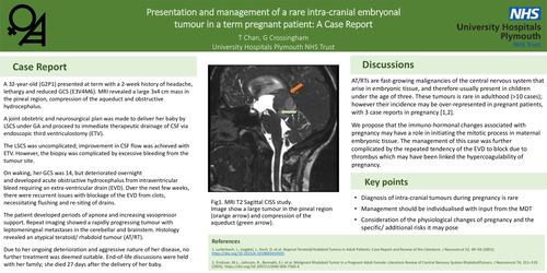
A 32-year-old (G2P1) presented at term with a 2-week history of headache, lethargy and reduced GCS (E3V4M6). MRI revealed a large 3x4 cm mass in the pineal region, compression of the aqueduct and obstructive hydrocephalus.
A joint obstetric and neurosurgical plan was made to deliver her baby by LSCS under GA and proceed to immediate therapeutic drainage of CSF via endoscopic third ventriculostomy (ETV).
The LSCS was uncomplicated; improvement in CSF flow was achieved with ETV. However, the biopsy was complicated by excessive bleeding from the tumour site.
On waking, her GCS was 14, but deteriorated overnight and developed acute obstructive hydrocephalus from intraventricular bleed requiring an extra-ventricular drain (EVD). Over the next few weeks, there were recurrent issues with blockage of the EVD from clots, necessitating flushing and re-siting of drains.
The patient developed periods of apnoea and increasing vasopressor support. Repeat imaging showed a rapidly progressing tumour with leptomeningeal metastases in the cerebellar and brainstem. Histology revealed an atypical teratoid/ rhabdoid tumour (AT/RT).
Due to her ongoing deterioration and aggressive nature of her disease, no further treatment was deemed suitable. End-of-life discussions were held with her family; she died 27 days after the delivery of her baby.
Discussions
AT/RTs are fast-growing malignancies of the central nervous system that arise in embryonic tissue, and therefore usually present in children under the age of three. These tumours is rare in adulthood (>10 cases); however their incidence may be over-represented in pregnant patients, with 3 case reports in pregnancy [1,2].
We propose that the immuno-hormonal changes associated with pregnancy may have a role in initiating the mitotic process in maternal embryonic tissue. The management of this case was further complicated by the repeated tendency of the EVD to block due to thrombus which may have been linked the hypercoagulability of pregnancy.
Key Points