301 posters, 50 videos, 13 topics, 13 sessions, 739 authors, 192 institutions
ePostersLive by SciGen Technologies S.A. All rights reserved.
ISMICS 2026 Annual Meeting
10 - 13 June, 2026 | Miami, Florida

D172
Intramyocardial LAD During MIDCAB: Staying The Course
Poster Presenter
Authors
Saad Hasan, Juan Umana-Pizano, Michael Javorski, Jeffrey Griffin, Robert Dowling, Geoffrey Answini, J. Michael Smith
Affiliations
Part of Topic
Coronary Revascularization
Video
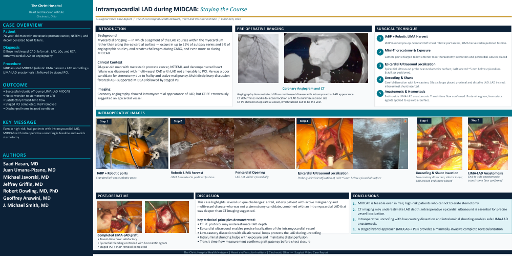
Objective: This video demonstrates a technique for treating an intramyocardial left anterior descending (LAD) artery during minimally-invasive direct coronary artery bypass (MIDCAB).
Methods: A 78-year-old man with metastatic prostate cancer presented with unstable angina and decompensated heart failure, work-up for which revealed NSTEMI and diffuse multi-vessel coronary artery disease involving the left main, LAD, left circumflex, and right coronary arteries. He was a poor candidate for sternotomy due to frailty and active malignancy. Following multidisciplinary discussion, the decision was made to proceed with intra-aortic balloon pump (IABP) assisted robotic left internal mammary artery (LIMA) to LAD bypass followed by staged percutaneous coronary intervention (PCI). The LAD had an intramyocardial appearance on angiography. CT PE protocol provided a misleadingly superficial appearance of the LAD.
Results: An IABP was inserted, and standard left chest robotic IMA harvest port access was established. The LIMA was harvested in pedicled fashion. The pericardium was opened, but only the ramus intermedius could be visualized epicardially. Heparin was given, and the LIMA was clipped, divided, and secured to the pericardium. The camera port was enlarged into a left anterior mini-thoracotomy, retractors were placed, and the LIMA was sprayed with papaverine. Pericardial sutures were placed, and an epicardial ultrasound probe was used to locate the LAD target. The epicardial surface was scanned across the left and right ventricles anteriorly, revealing the LAD target about 5mm below the surface. An epicardial stabilizer was placed, and careful dissection was performed with low cautery. Multiple passes of the ultrasound probe and repositioning of the epicardial stabilizer were required, as well as numerous attempts at dissection. Ultimately, silastic vessel loops were placed deeply proximal and distal to the identified location of the LAD target. The epicardial stabilizer position was optimized. The LAD was finally incised, and a shunt was inserted.The anastomosis was performed in the usual end-to-side fashion. Transit-time flow measurement was satisfactory. Heparin was reversed with protamine. The epicardial surface was packed while protamine was given. Hemostatic agents were subsequently used to eliminate epicardial bleeding.
Conclusions: The patient was ultimately discharged in good condition following staged PCI and IABP removal.