301 posters, 50 videos, 13 topics, 13 sessions, 734 authors, 193 institutions
ePostersLive by SciGen Technologies S.A. All rights reserved.
ISMICS 2026 Annual Meeting
10 - 13 June, 2026 | Miami, Florida

D150
Robot-Assisted Repair Of Massive Type IV Hiatal Hernia
Poster Presenter
Part of Topic
Thoracic Surgery � Miscellaneous
Video
BACKGROUND
Type IV hiatal hernias are the rarest and most complex form of paraesophageal
hernia, involving herniation of multiple intra-abdominal organs — including the entire
stomach, transverse colon, and omentum — into the mediastinum.
Surgical repair is technically demanding due to:
• Extensive mediastinal dissection required
• Risk of bilateral pleural violation
• Need for adequate esophageal length
• Minimizing operative morbidity
Robotic-assisted approaches may facilitate the precision dissection and suturing required in these highly complex cases.
CASE PRESENTATION
46-Year-Old Male — Type IV Hiatal Hernia
PMH: Asthma, GERD, chronic cough | Referred by GI | No prior surgery
Preoperative workup:
• Esophagogastroduodenoscopy (EGD) with biopsy
• Barium esophagram
• Esophageal manometry
• CT chest — coronal view shown below
OBJECTIVES
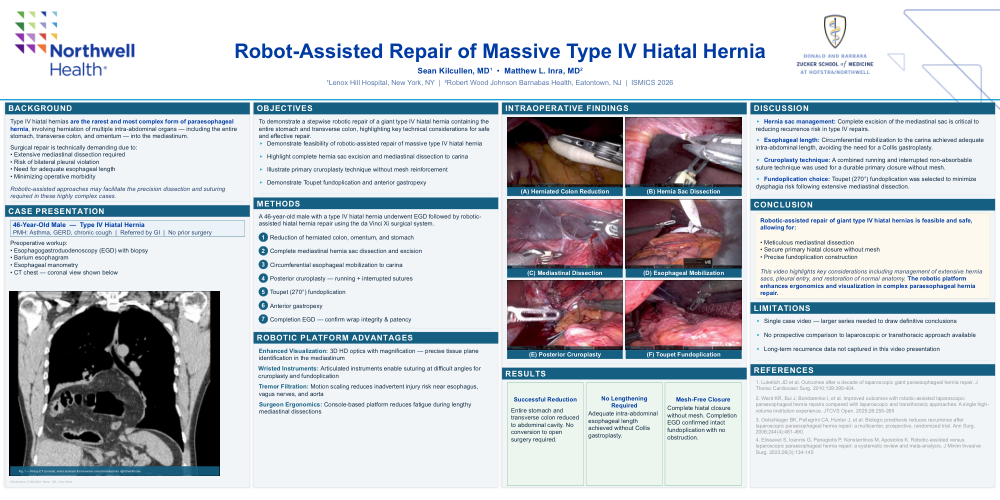
To demonstrate a stepwise robotic repair of a giant type IV hiatal hernia containing the
entire stomach and transverse colon, highlighting key technical considerations for safe
and effective repair.
▸Demonstrate feasibility of robotic-assisted repair of massive type IV hiatal hernia
▸Highlight complete hernia sac excision and mediastinal dissection to carina
▸Illustrate primary cruroplasty technique without mesh reinforcement
▸Demonstrate Toupet fundoplication and anterior gastropexy
METHODS
A 46-year-old male with a type IV hiatal hernia underwent EGD followed by robotic-
assisted hiatal hernia repair using the da Vinci Xi surgical system.
1 Reduction of herniated colon, omentum, and stomach
2 Complete mediastinal hernia sac dissection and excision
3 Circumferential esophageal mobilization to carina
4 Posterior cruroplasty — running + interrupted sutures
5 Toupet (270°) fundoplication
6 Anterior gastropexy
7 Completion EGD — confirm wrap integrity & patency
ROBOTIC PLATFORM ADVANTAGES
Enhanced Visualization: 3D HD optics with magnification — precise tissue plane
identification in the mediastinum
Wristed Instruments: Articulated instruments enable suturing at difficult angles for
cruroplasty and fundoplication
Tremor Filtration: Motion scaling reduces inadvertent injury risk near esophagus,
vagus nerves, and aorta
Surgeon Ergonomics: Console-based platform reduces fatigue during lengthy
mediastinal dissections
RESULTS
Successful Reduction
Entire stomach and transverse colon reduced to abdominal cavity. No conversion to open surgery required.
No Lengthening
Required
Adequate intra-abdominal esophageal lengthachieved without Collisngastroplasty.
Mesh-Free Closure
Complete hiatal closurenwithout mesh. Completion EGD confirmed intact fundoplication with no obstruction.
DISCUSSION
▸Hernia sac management: Complete excision of the mediastinal sac is critical to
reducing recurrence risk in type IV repairs.
▸Esophageal length: Circumferential mobilization to the carina achieved adequate
intra-abdominal length, avoiding the need for a Collis gastroplasty.
▸Cruroplasty technique: A combined running and interrupted non-absorbable
suture technique was used for a durable primary closure without mesh.
▸Fundoplication choice: Toupet (270°) fundoplication was selected to minimize
dysphagia risk following extensive mediastinal dissection.
CONCLUSION
Robotic-assisted repair of giant type IV hiatal hernias is feasible and safe,
allowing for:
• Meticulous mediastinal dissection
• Secure primary hiatal closure without mesh
• Precise fundoplication construction
This video highlights key considerations including management of extensive hernia
sacs, pleural entry, and restoration of normal anatomy. The robotic platform
enhances ergonomics and visualization in complex paraesophageal hernia
repair.
LIMITATIONS
▸Single case video — larger series needed to draw definitive conclusions
▸No prospective comparison to laparoscopic or transthoracic approach available
▸Long-term recurrence data not captured in this video presentation
REFERENCES
1. Luketich JD et al. Outcomes after a decade of laparoscopic giant paraesophageal hernia repair. J
Thorac Cardiovasc Surg. 2010;139:395-404.
2. Ward KR, Bui J, Bondarenko I, et al. Improved outcomes with robotic-assisted laparoscopic
paraesophageal hernia repairs compared with laparoscopic and transthoracic approaches: A single high-
volume institution experience. JTCVS Open. 2025;26:255-265
3. Oelschlager BK, Pellegrini CA, Hunter J, et al. Biologic prosthesis reduces recurrence after
laparoscopic paraesophageal hernia repair: a multicenter, prospective, randomized trial. Ann Surg.
2006;244(4):481-490.
4. Elissavet S, Ioannis G, Panagiotis P, Konstantinos M, Apostolos K. Robotic-assisted versus
laparoscopic paraesophageal hernia repair: a systematic review and meta-analysis. J Minim Invasive
Surg. 2023;26(3):134-145