301 posters, 50 videos, 13 topics, 13 sessions, 739 authors, 192 institutions
ePostersLive by SciGen Technologies S.A. All rights reserved.
ISMICS 2026 Annual Meeting
10 - 13 June, 2026 | Miami, Florida

D104
Diagnostic Uncertainty Of A Pulsatile Mass After Transapical Beating-Heart Mitral Valve Repair
Part of Topic
Valve � Surgical Valves
Title: Diagnostic Uncertainty of a Pulsatile Mass After Transapical Beating-Heart Mitral Valve Repair
Authors: Navneet Kang, BSc¹; Amy Brown, MD, MSc, MPH²; Toshiro Sembo, MD²; Bob Kiaii, MD²; William D. T. Kent, MD, MSc²; Corey Adams, MD, MSc²
Affiliations: ¹ Ottawa Heart Institute, Ottawa, ON, Canada ² Department of Cardiac Sciences, Libin Cardiovascular Institute, University of Calgary, Calgary, AB, Canada
Background: Mitral valve repair is the preferred treatment for degenerative mitral regurgitation (MR), but conventional surgery requires sternotomy and cardiopulmonary bypass (CPB). Transcatheter edge-to-edge repair is an off-pump alternative but achieves optimal results only in select anatomy, leaving many high-risk patients without an ideal option. Transapical beating-heart mitral valve repair is a minimally invasive, off-pump technique performed through a left mini-thoracotomy, placing artificial chordae under transesophageal echocardiographic guidance. Complications of the transapical access tract remain incompletely characterized — a delayed pulsatile mass may reflect a structural complication or an indolent infection.
Case Presentation: An 85-year-old man with symptomatic severe degenerative MR underwent off-pump transapical beating-heart mitral valve repair via a left mini-thoracotomy. Six months later he presented with two enlarging, pulsatile subcutaneous masses at the thoracotomy site. He was afebrile and hemodynamically stable, with normal inflammatory markers and no leukocytosis.
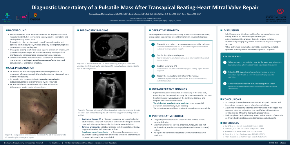
Diagnostic Imaging: Contrast-enhanced CT demonstrated a ~5 cm rim-enhancing peri-apical collection abutting the left ventricular apex, with two further collections tracking into the left chest wall; the myocardium–collection interface was indistinct. Targeted ultrasound showed a bilobed anechoic collection contacting the left ventricle; Doppler showed no definitive internal flow. Imaging remained inconclusive — a thrombosed pseudoaneurysm could not be distinguished from an infected collection, and ventricular communication could not be excluded.
Operative Strategy — Safe Re-Entry Pathway: Because pseudoaneurysm rupture during re-entry could not be excluded, the operation was planned around the higher-risk structural diagnosis. (1) Diagnostic uncertainty — pseudoaneurysm cannot be excluded; equivocal CT and ultrasound, the LV–collection interface is indistinct and ventricular communication is possible. (2) Plan for the higher-risk diagnosis; treat as a structural complication until proven otherwise to reduce risk of harm to patient. (3) Establish peripheral CPB; femoral arterial and venous cannulation, bypass running before the chest is opened. (4) Reopen the thoracotomy only after CPB is running; converts an unpredictable, potentially lethal re-entry into a controlled, protected operation.
Intraoperative Findings: Exploration revealed a loculated abscess cavity in the chest wall, extending into the pericardium along the prior transapical access tract. Purulent material was evacuated; the cavity was debrided and irrigated and adhesions were lysed. The pledgetted apical entry site was intact — no myocardial disruption, pseudoaneurysm, or bleeding. The patient was weaned from cardiopulmonary bypass uneventfully.
Postoperative Course: The postoperative course was uncomplicated and the patient remained afebrile. Specimens underwent aerobic, anaerobic, fungal, and acid-fast bacillus culture, with broad-range polymerase chain reaction (PCR) testing. No organisms were identified; broad-spectrum antibiotics were continued.
Discussion: Late thoracotomy-site abnormalities after transapical access can closely mimic a left ventricular pseudoaneurysm. Altered postoperative anatomy degrades imaging certainty — multimodality imaging may remain inconclusive despite a complete workup. When a structural complication cannot be confidently excluded, operative planning should assume the higher-risk diagnosis.
Key Takeaways: (1) When imaging is inconclusive, plan for the worst-case diagnosis — equivocal findings after transapical access require operative caution. (2) Establish CPB by peripheral cannulation before re-entry — convert an unpredictable re-entry into a controlled, reproducible operation. (3) A reproducible framework that prioritizes patient safety — adoptable by any team facing late post-transapical thoracotomy-site abnormalities.
Conclusions: As transapical access becomes more widely adopted, clinicians will increasingly encounter access-related complications. A pulsatile thoracotomy-site mass after transapical mitral repair may represent infection rather than structural failure, although these entities may be difficult to distinguish preoperatively. Early peripheral cardiopulmonary bypass before re-entry offers a safe and reproducible strategy when diagnostic uncertainty exists.
Figure Legends: Figure 1. Two pulsatile subcutaneous masses at the left thoracotomy site, six months after transapical repair. Figure 2. Contrast-enhanced CT. Rim-enhancing peri-apical collection abutting the left ventricular apex (asterisk); two collections extend into the left chest wall (arrows). Figure 3. Targeted ultrasound. Bilobed anechoic collection tracking deep to the chest wall and contacting the left ventricle; Doppler limited by motion artifact.
Keywords: transapical mitral valve repair, beating-heart mitral repair, artificial chordae, pseudoaneurysm, thoracotomy-site infection, diagnostic uncertainty, cardiopulmonary bypass, peripheral cannulation, mini-thoracotomy, mitral regurgitation, case report.
Disclosures: The authors report no conflicts of interest. Funding: none. Written informed consent for publication was obtained from the patient.