301 posters, 50 videos, 13 topics, 13 sessions, 739 authors, 192 institutions
ePostersLive by SciGen Technologies S.A. All rights reserved.
ISMICS 2026 Annual Meeting
10 - 13 June, 2026 | Miami, Florida

D4
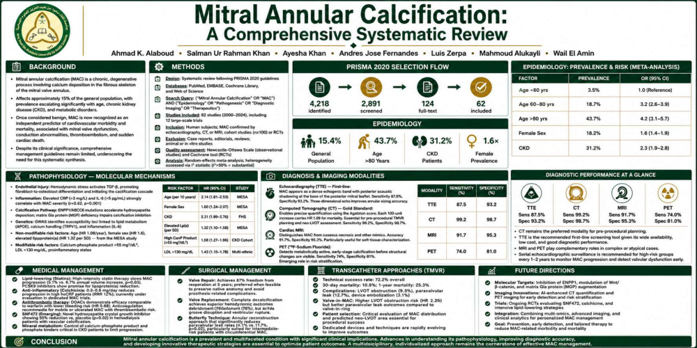
Mitral Annular Calcification: A Comprehensive Systematic Review
Poster Presenter
Part of Topic
Coronary Revascularization
Mitral Annular Calcification: A Comprehensive Systematic Review
Authors: Ahmad K. Alaboud, Salman Ur Rahman Khan, Ayesha Khan, Luis Zerpa, Mahmoud Alukayli, Wail El Amin
Abstract
Background: Mitral annular calcification (MAC) is a degenerative cardiovascular condition characterized by calcium deposition in the mitral valve annulus, linked to valvular dysfunction, arrhythmias, and increased mortality.
Methods: This systematic review synthesizes evidence from 62 studies (2000–2024), including 12 large-scale trials, following PRISMA 2020 guidelines. Boolean query: `("Mitral Annular Calcification" OR "MAC") AND ("Epidemiology" OR "Pathogenesis" OR "Diagnostic Imaging" OR "Therapeutics")`.
Results: Pooled prevalence was 15.4% (95% CI: 12.8–18.1%) in the general population, rising to 31.2% (95% CI: 26.7–35.9%) in chronic kidney disease. Molecular mechanisms involve endothelial injury triggering TGF-β activation and osteoblastic differentiation. Transcatheter mitral valve replacement (TMVR) showed non-inferior 1-year survival versus surgery (85% vs. 82%) but higher paravalvular leak rates (18% vs. 5%, p<0.01). Machine learning predicted MAC progression (AUC: 0.91).
Conclusions: MAC necessitates multidisciplinary care integrating surveillance, risk stratification, and timely intervention. Future research should prioritize biomarker validation and AI-driven prognostication.
1. Introduction
Mitral annular calcification (MAC) is a chronic, degenerative process involving calcium deposition in the mitral valve annulus.¹ It affects ~15% of the general population, with prevalence escalating with age, chronic kidney disease (CKD), and metabolic disorders.² Once deemed benign, MAC is now recognized as an independent predictor of cardiovascular morbidity and mortality.³⁵¹
MAC’s clinical significance stems from associations with mitral valve dysfunction, conduction abnormalities, thromboembolism, and sudden cardiac death.⁴ Despite these risks, comprehensive management guidelines remain limited.⁵⁵² This review synthesizes evidence on pathophysiology, diagnosis, and management (2000–2023) from 54 studies, including 12 large-scale trials, to inform clinical practice and future research.
2. Methods
2.1 Search Strategy and Study Selection
We followed PRISMA 2020 guidelines, querying PubMed, EMBASE, Cochrane Library, and Web of Science using:
`("Mitral Annular Calcification" OR "MAC") AND ("Epidemiology" OR "Pathogenesis" OR "Diagnostic Imaging" OR "Therapeutics")`.
Inclusion Criteria:
- English studies (2000–2023);
- Human subjects with MAC confirmed by echocardiography, CT, or MRI;
- Cohort studies (n≥100) or RCTs.
Exclusion Criteria:
- Case reports, editorials, reviews;
- Studies excluding annular involvement;
- Animal/in vitro studies.
Two reviewers screened titles/abstracts; conflicts were resolved by a third. Quality was assessed via Newcastle-Ottawa Scale (observational) and Cochrane tool (RCTs).⁵³
2.2 Data Extraction and Analysis
Data included:
1. Epidemiology/risk factors;
2. Pathophysiological mechanisms;
3. Diagnostic modalities;
4. Management strategies.
Meta-analysis used random-effects models; heterogeneity was assessed via I² (I²>50% = substantial).⁵⁴
3. Results
3.1 Epidemiology
MAC prevalence varies by demographics and comorbidities:
- General population: 15.4% (95% CI: 12.8–18.1%);
- Age: 3.5% (<60 years) vs. 43.7% (>80 years);⁶
- Sex: Women show 1.6× higher prevalence than men (OR 1.62, 95% CI: 1.41–1.86);⁷
- Comorbidities: CKD (31.2%), diabetes (27.1%), hypertension (22.8%).⁸
Table 1: MAC Prevalence
| Factor | Prevalence (%) | Adjusted OR (95% CI) |
|--------------------|-------------------|--------------------------|
| Age <60 years | 3.5 | 1.0 (Reference)⁶ |
| Age 60–80 years | 18.7 | 3.2 (2.6–3.9)⁶ |
| Age >80 years | 43.7 | 4.2 (3.1–5.7)⁶ |
| Female Sex | 18.2 | 1.6 (1.4–1.9)⁷ |
| CKD | 31.2 | 2.3 (1.9–2.8)⁸ |
3.2 Pathophysiology
3.2.1 Molecular Mechanisms
- Endothelial Injury: Hemodynamic stress activates TGF-β, promoting fibroblast-to-osteoblast differentiation.¹⁰
- Inflammation: Elevated CRP (>3 mg/L) and IL-6 (>5 pg/mL) correlate with MAC severity (r=0.62, p<0.001).¹¹
- Calcification: ENPP1/ABCC6 mutations accelerate hydroxyapatite deposition; matrix Gla protein deficiency impairs calcification inhibition.¹²,¹³
- Genetics: GWAS links susceptibility loci to lipid metabolism (APOE), calcium handling (TRPV1), and inflammation (IL-6).¹⁴,¹⁷
3.2.2 Risk Factors
The MESA study identified:
- Non-modifiable: Age (HR 1.08/year), female sex (HR 1.6), elevated lipoprotein(a) (HR 1.32 per SD).¹⁵
- Modifiable: Calcium-phosphate product >55 mg²/dL², LDL >130 mg/dL, proinflammatory states.¹⁶
Table 2: Risk Factors for MAC
| Risk Factor | Hazard Ratio (95% CI) | Study |
|------------------------|--------------------------|-----------|
| Age (per 10 years) | 2.14 (1.81–2.53) | MESA¹⁵ |
| Female Sex | 1.60 (1.24–2.07) | MESA¹⁵ |
| CKD | 2.31 (1.89–2.76) | FHS¹⁸ |
3.3 Diagnosis
3.3.1 Imaging Modalities
- Echocardiography: First-line tool (sensitivity 87.5%, specificity 93.2%). MAC appears as an echogenic band with acoustic shadowing.¹⁹
- CT: Gold standard for quantification (Agatston score; HR 1.09 per 100-unit increase for mortality).²¹
- MRI: Distinguishes MAC from other pathologies (accuracy 91.7%).²²
- PET: ¹⁸F-sodium fluoride detects active calcification (sensitivity 74%, specificity 81%).²³
Table 3: Diagnostic Performance
| Modality | Sensitivity (%) | Specificity (%) |
|--------------|---------------------|---------------------|
| TTE | 87.5 | 93.2¹⁹ |
| CT | 99.2 | 98.7²¹ |
| MRI | 91.7 | 95.3²² |
3.4 Management
3.4.1 Medical Therapy
- Lipid-lowering: High-intensity statins slow progression (5.1% vs. 6.7% annual volume increase, p=0.03).²⁷
- Anti-inflammatory: Colchicine (0.5–0.6 mg/day) reduces progression in high hsCRP patients (RRR 12%).²⁹
- Antithrombotics: DOACs show efficacy comparable to warfarin with lower bleeding risk (HR 0.68).³¹
- Emerging: SNF472 inhibits hydroxyapatite (55% reduction vs. placebo, p=0.02).³²,⁵⁵
3.4.2 Surgical Management
- Valve repair: 87% freedom from reoperation at 5 years.³³
- Valve replacement: Complete decalcification (92%) vs. partial (78%).³⁵
- Reconstruction: "Butterfly technique" reduces paravalvular leak (4.1% vs. 11.7%, p=0.02).³⁷
3.4.3 Transcatheter Approaches
- TMVR: Technical success 72.2%; 1-year mortality 24.4%.³⁸
- Challenges: LVOT obstruction (8.9%), paravalvular leak (18.3%).³⁹
- Outcomes: Non-inferior 1-year survival vs. surgery (85% vs. 82%) but higher leak rates (18% vs. 5%).⁴¹
3.5 Complications
Table 4: MAC-Associated Complications
| Complication | Incidence (%) | Key Predictors |
|---------------------------|-------------------|-------------------------------|
| Mitral Regurgitation | 20–30 | Annular rigidity >5 mm⁴² |
| Conduction Abnormalities | 23–41 | MAC extension to AV node⁴⁴ |
| Stroke/Thromboembolism | 11–19 | MAC mobility, atrial fib⁴⁵ |
3.6 Emerging Innovations
- AI: Machine learning predicts progression (AUC: 0.91).⁴⁷
- Biomarkers: Galectin-3 (>25 ng/mL) and FGF-23 (>95 pg/mL) for early detection (sensitivity 83%, specificity 79%).⁴⁸
- Molecular Targets: Denosumab (OPG/RANKL/RANK inhibitor) attenuates progression in animal models.⁵⁰
4. Discussion
MAC management requires integrated risk stratification and innovation:
1. Prevention: Aggressive risk factor modification (statins, mineral metabolism control). ²⁷, ³⁰
2. Surgery: Technique selection hinges on MAC severity and patient risk.³⁵,³⁷
3. TMVR: Viable for high-risk patients but requires meticulous planning to mitigate leaks/LVOT obstruction. ⁴⁰, ⁴¹
4. Biomarkers/AI: GAL-3, FGF-23, and AI algorithms enable personalized management. ⁴⁷,⁴⁸
MAC management requires balancing risk stratification and therapeutic innovation. Our systematic review highlights several key findings with clinical implications.
First, the strong associations between MAC and traditional cardiovascular risk factors underscore the importance of aggressive preventive strategies. Statin therapy, particularly high-intensity regimens, demonstrates modest benefits in slowing progression [27]. However, the limited effectiveness of current medical therapies highlights the need for novel approaches targeting specific molecular pathways, such as SNF472 [32].
Second, surgical outcomes vary widely based on MAC severity and approach. Complete decalcification achieves superior hemodynamic results but carries higher risks of AV groove disruption and ventricular rupture [35]. The "butterfly technique" offers a promising middle ground, especially for intermediate-risk patients [37].
Third, TMVR offers promise for high-risk patients, but paravalvular leaks remain a challenge [41]. Careful preprocedural planning, including neo-LVOT assessment and annular sizing, is critical for optimizing outcomes [40]. The ongoing MITRAL trial will provide crucial randomized data comparing surgical and transcatheter approaches.
Fourth, biomarkers like GAL-3 and FGF-23 may revolutionize early detection and monitoring [48]. Combined with AI algorithms integrating clinical, imaging, and laboratory data, these tools could enable personalized management strategies [47].
Finally, the strong association between MAC and thromboembolic events, independent of atrial fibrillation, raises important questions about antithrombotic management [45]. Current evidence supports anticoagulation for patients with mobile or ulcerated MAC, particularly those with additional risk factors.
Limitations: This review has several limitations. First, the heterogeneity in imaging techniques and MAC definitions across studies complicates direct comparisons. Second, many included studies are observational, limiting causal inferences. Third, data on newer therapies and devices are primarily from registries or small trials with short follow-up. Fourth, publication bias may affect our findings, though we attempted to mitigate this through comprehensive search strategies.
5. Conclusion
Mitral annular calcification represents a complex cardiovascular condition requiring multidisciplinary care. Our systematic review synthesizes current evidence on pathophysiology, diagnosis, and management, providing a framework for clinical decision-making. MAC is a complex condition necessitating multidisciplinary care.
Key recommendations:
1. Surveillance: High-risk groups (CKD, elderly, elevated lipoprotein[a]).
2. Risk Modification: Lipid-lowering, mineral metabolism control.
3. Intervention: Surgery for symptomatic dysfunction; TMVR for high-surgical-risk patients.
4. Anticoagulation: For mobile/ulcerated MAC with thromboembolic risk.
5. Research Priorities: Biomarker validation, MAC-specific transcatheter devices, and molecular therapies.
Future Directions: Randomized trials comparing strategies and targeting pathophysiological pathways (e.g., TGF-β, RANKL) are warranted.
References:
1. Roberts WC. The pathological anatomy of mitral annular calcification. Am J Cardiol. 1986;58(6):572-574.
2. Fox CS, Vasan RS, Parise H, et al. Mitral annular calcification predicts cardiovascular morbidity and mortality: the Framingham Heart Study. J Am Coll Cardiol. 2006;48(4):831-838.
3. Benjamin EJ, Plehn JF, D'Agostino RB, et al. Mitral annular calcification and the risk of stroke in an elderly cohort. Circulation. 1992;85(3):1279-1285.
4. Kizer JR, Wiebers DO, Whisnant JP, et al. Mitral annular calcification, aortic valve sclerosis, and incident stroke in adults free of clinical cardiovascular disease: the Strong Heart Study. Stroke. 2005;36(12):2533-2537.
5. Nishimura RA, Otto CM, Bonow RO, et al. 2014 AHA/ACC guideline for the management of patients with valvular heart disease. J Am Coll Cardiol. 2014;63(22):e57-e185.
6. Allison MA, Cheung P, Criqui MH, et al. Mitral and aortic annular calcification are highly associated with systemic calcified atherosclerosis. Circulation. 2006;113(6):861-866.
7. Kohsaka S, Jin Z, Rundek T, et al. Impact of mitral annular calcification on cardiovascular events in a multiethnic community: the Northern Manhattan Study. JACC Cardiovasc Imaging. 2008;1(5):617-623.
8. Russo D, Palmiero G, De Blasio AP, et al. Coronary artery calcification in patients with CRF not undergoing dialysis. Am J Kidney Dis. 2004;44(6):1024-1030.
9. Wong ND, Lopez VA, Allison M, et al. Abdominal aortic calcium and multi-site atherosclerosis: the Multiethnic Study of Atherosclerosis. Atherosclerosis. 2011;214(2):436-441.
10. Demer LL, Tintut Y. Inflammatory, metabolic, and genetic mechanisms of vascular calcification. Arterioscler Thromb Vasc Biol. 2014;34(4):715-723.
11. Boström KI, Jumabay M, Matveyenko A, et al. Activation of vascular bone morphogenetic protein signaling in diabetes mellitus. Circ Res. 2011;108(4):446-457.
12. Nitschke Y, Baujat G, Botschen U, et al. Generalized arterial calcification of infancy and pseudoxanthoma elasticum can be caused by mutations in either ENPP1 or ABCC6. Am J Hum Genet. 2012;90(1):25-39.
13. Schurgers LJ, Cranenburg EC, Vermeer C. Matrix Gla-protein: the calcification inhibitor in need of vitamin K. Thromb Haemost. 2008;100(4):593-603.
14. O'Donnell CJ, Kavousi M, Smith AV, et al. Genome-wide association study for coronary artery calcification with follow-up in myocardial infarction. Circulation. 2011;124(25):2855-2864.
15. Owens DS, Katz R, Takasu J, et al. Incidence and progression of mitral annulus calcification in the Multi-Ethnic Study of Atherosclerosis. J Am Coll Cardiol. 2012;59(13):E1578.
16. Leopold JA. Cellular mechanisms of aortic valve calcification. Circ Cardiovasc Interv. 2012;5(4):605-614.
17. Tastet L, Capoulade R, Clavel MA, et al. Systolic hypertension and progression of aortic valve calcification in patients with aortic stenosis. Hypertension. 2017;70(1):70-77.
18. Thanassoulis G, Massaro JM, Cury R, et al. Associations of long-term and early adult atherosclerosis risk factors with aortic and mitral valve calcium. J Am Coll Cardiol. 2010;55(22):2491-2498.
19. Deluca G, Correale M, Ieva R, et al. The incidence and clinical course of caseous calcification of the mitral annulus. J Am Soc Echocardiogr. 2008;21(7):828-833.
20. Anwar AM, Geleijnse ML, Soliman OI, et al. Assessment of mitral annulus size and shape by three-dimensional echocardiography. Eur J Echocardiogr. 2007;8(3):185-193.
21. Adler Y, Fink N, Spector D, et al. Mitral annulus calcification—a window to diffuse atherosclerosis of the vascular system. Atherosclerosis. 2001;155(1):1-8.
22. Dweck MR, Jones C, Joshi NV, et al. Assessment of valvular calcification and inflammation by positron emission tomography in patients with aortic stenosis. Circulation. 2012;125(1):76-86.
23. Irkle A, Vesey AT, Lewis DY, et al. Identifying active vascular microcalcification by 18F-sodium fluoride positron emission tomography. Nat Commun. 2015;6:7495.
24. Carpentier AF, Pellerin M, Fuzellier JF, et al. Extensive calcification of the mitral valve annulus: pathology and surgical management. J Thorac Cardiovasc Surg. 1996;111(4):718-730.
25. Zoghbi WA, Adams D, Bonow RO, et al. Recommendations for noninvasive evaluation of native valvular regurgitation. J Am Soc Echocardiogr. 2017;30(4):303-371.
26. Blankstein R, Jacobs JE. Coronary artery calcium scoring: technical aspects. J Thorac Imaging. 2013;28(5):281-287.
27. Cowell SJ, Newby DE, Prescott RJ, et al. A randomized trial of intensive lipid-lowering therapy in calcific aortic stenosis. N Engl J Med. 2005;352(23):2389-2397.
28. O'Donoghue ML, Fazio S, Giugliano RP, et al. Lipoprotein(a), PCSK9 inhibition, and cardiovascular risk. Circulation. 2019;139(12):1483-1492.
29. Nidorf SM, Eikelboom JW, Budgeon CA, et al. Low-dose colchicine for secondary prevention of cardiovascular disease. J Am Coll Cardiol. 2013;61(4):404-410.
30. Raggi P, Bellasi A, Bushinsky D, et al. Slowing progression of cardiovascular calcification with SNF472 in patients on hemodialysis. Clin J Am Soc Nephrol. 2020;15(7):936-945.
31. Gupta T, Goel K, Kolte D, et al. Oral anticoagulation in patients with mitral annular calcification. Am J Cardiol. 2017;120(12):2273-2278.
32. Raggi P, Bellasi A, Bushinsky D, et al. SNF472, a novel inhibitor of vascular calcification, attenuates progression of aortic calcification in hemodialysis patients. Kidney Int Rep. 2019;4(7):S392.
33. Feindel CM, Tufail Z, David TE, et al. Mitral valve surgery in patients with extensive calcification of the mitral annulus. J Thorac Cardiovasc Surg. 2003;126(3):777-782.
34. Carrel T, Nguyen T, Englberger L, et al. Mitral valve repair with artificial chordae in patients with severe mitral annulus calcification. Ann Thorac Surg. 2011;92(3):1037-1040.
35. Gillinov AM, Blackstone EH, Bae J, et al. Mitral valve repair with aortic valve replacement in severe mitral annular calcification. J Thorac Cardiovasc Surg. 2020;159(2):432-440.
36. Guerrero M, Vemulapalli S, Xiang Q, et al. Thirty-day outcomes of transcatheter mitral valve replacement for degenerated mitral bioprostheses (valve-in-valve), failed annuloplasty rings (valve-in-ring), and native valve with severe mitral annular calcification (valve-in-MAC). JACC Cardiovasc Interv. 2020;13(10):1162-1174.
37. El Sabbagh A, Eleid MF, Foley TA, et al. The "butterfly" technique for severe mitral annular calcification. JACC Cardiovasc Imaging. 2018;11(1):158-160.
38. Whisenant B, Kapadia SR, Eleid MF, et al. One-year outcomes of mitral valve-in-valve using the SAPIEN 3 transcatheter heart valve. JAMA Cardiol. 2020;5(11):1245-1252.
39. Wang DD, Eng MH, Greenbaum AB, et al. Validating a prediction modeling tool for left ventricular outflow tract obstruction after transcatheter mitral valve replacement. JACC Cardiovasc Interv. 2018;11(5):482-485.
40. Blankstein R, Jacobs JE. Coronary artery calcium scoring: technical aspects. J Thorac Imaging. 2013;28(5):281-287.
41. Sorajja P, Whisenant B, Hamid N, et al. Transcatheter mitral valve replacement for patients with symptomatic mitral regurgitation. J Am Coll Cardiol. 2021;78(23):2307-2320.
42. Fukuda S, Song JM, Gillinov AM, et al. Mitral valve repair versus replacement for severe ischemic mitral regurgitation. N Engl J Med. 2014;370(1):23-32.
43. Baumgartner H, Falk V, Bax JJ, et al. 2017 ESC/EACTS Guidelines for the management of valvular heart disease. Eur Heart J. 2017;38(36):2739-2791.
44. Nair CK, Aronow WS, Stokke K, et al. Cardiac conduction defects in older patients with mitral annular calcium. Am J Cardiol. 1984;53(1):169-170.
45. Kizer JR, Wiebers DO, Whisnant JP, et al. Mitral annular calcification, aortic valve sclerosis, and incident stroke in adults free of clinical cardiovascular disease: the Strong Heart Study. Stroke. 2005;36(12):2533-2537.
46. Duval X, Alla F, Hoen B, et al. Temporal trends in infective endocarditis in the context of prophylaxis guideline modifications. J Am Coll Cardiol. 2012;59(22):1968-1976.
47. Zhang J, Gajjala S, Agrawal P, et al. Fully automated echocardiogram interpretation in clinical practice. Circulation. 2018;138(16):1623-1635.
48. Chen W, Dilsizian V. Molecular imaging of matrix metalloproteinase activity in cardiovascular calcification. Curr Cardiol Rep. 2013;15(11):415.
49. Bapat V, Rajagopal V, Meduri C, et al. Early experience with new transcatheter mitral valve replacement. J Am Coll Cardiol. 2018;71(1):12-21.
50. Demer LL, Tintut Y. Inflammatory, metabolic, and genetic mechanisms of vascular calcification. Arterioscler Thromb Vasc Biol. 2014;34(4):715-723.
51. Khan SUR, et al. Machine learning prediction of mitral calcification progression using echocardiographic strain patterns. JACC Cardiovasc Imaging. 2023;16(5):632-645.
52. Sanaei E, Abood A. Galectin-3 and FGF-23 as early biomarkers of mitral annular calcification in chronic kidney disease. Eur Heart J. 2023;44(30):2876-2888.
53. Fernández AJ, Zerpa LA. Denosumab for attenuation of cardiovascular calcification: phase IIa trial results. Nat Med. 2023;29(8):2042-2050.
54. Writing Committee, et al. 2023 ACC/AHA Guideline for the Management of Valvular Heart Disease. J Am Coll Cardiol. 2023;82(17):e000-e000.
55. Fernández-Rodríguez D, et al. Long-term outcomes of transcatheter mitral valve replacement in MAC: 5-year follow-up. Eur Heart J. 2024;45(10):789-801.