293 posters, 56 videos, 24 topics, 13 sessions, 658 authors, 215 institutions
ePostersLive by SciGen Technologies S.A. All rights reserved.
ISMICS 2025 Annual Meeting
28 - 31 May, 2025 | Istanbul, Turkey

D136
Long-standing Undetected Endobronchial Tumor Leading To Recurrent Respiratory Infections And Segmental Destruction - Anatomic Segmentectomy
Poster Presenter
Authors
Part of Topic
Thoracic Surgery – Lung
Long-standing Undetected Endobronchial Tumor Leading To Recurrent Respiratory Infections And Segmental Destruction - Anatomic Segmentectomy
Introduction and Purpose:
Although endobronchial lesions often present with non-specific symptoms, these symptoms warrant further investigation, which can facilitate early diagnosis, especially in cases of recurrent post-obstructive lobar or segmental pneumonia. Recurrent segmental pneumonia can lead to bronchiectasis, and in advanced stages, concomitant segmental destruction and fibrosis.
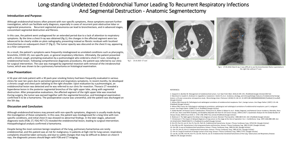
In this case, the patient went undiagnosed for an extended period due to a lack of attention to respiratory symptoms. By the time a chest X-ray was obtained (fig.1), the changes in the affected segment were too advanced to be clearly visible on plain radiography, presenting instead as fibrotic residuals with localized bronchiectasis on subsequent chest CT (fig.2). The tumor opacity was obscured on the chest X-ray, appearing as a hilar component.
As a result, the patient's symptoms were frequently misdiagnosed as unrelated conditions such as pharyngitis, bronchitis, COVID-19, non-specific pain, or general respiratory infections. Ultimately, the patient presented with a chronic cough, prompting evaluation by a pulmonologist who ordered a chest CT scan, revealing an endobronchial lesion. Following comprehensive diagnostic procedures, the patient was referred to our clinic for surgical intervention. The case was managed by segmental resection with removal of the intrabronchial tumor, which was shown to be a pulmonary hamartoma on histological examination.
Case Presentation:
A 56-year-old male patient with a 30 pack-year smoking history had been frequently evaluated in various clinics for over ten years due to persistent general and respiratory symptoms. In recent months, he developed a chronic cough and chest pain radiating to the right shoulder. Upon evaluation by a pulmonologist, an endobronchial lesion was detected and he was referred to our clinic for resection. Thoracic CT revealed a hyperdense lesion in the posterior segmental bronchus of the right upper lobe, along with segmental destruction. After preoperative evaluations, the affected segment of the right upper lobe was resected. During surgery, the tumor was excised together with the segmental bronchus, and histological examination confirmed it to be a hamartoma. The postoperative course was uneventful, and the patient was discharged on the 5th day.
Discussion and Conclusion:
Although endobronchial lesions may present with non-specific symptoms, diagnosis is usually made during the investigation of these complaints. In this case, the patient was misdiagnosed for a long time with non-specific conditions, and initial chest X-rays showed no abnormal findings. In the later stages, advanced diagnostic tools (FOB, CT, FDG-PET-CT) revealed the endobronchial lesion, leading to surgical intervention. Histopathological analysis confirmed a hamartoma.
Despite being the most common benign neoplasm of the lung, pulmonary hamartomas are rarely endobronchial, and this patient was at risk for malignancy. In patients at high risk for lung cancer, respiratory complaints should be taken seriously, and due to subtle changes that may be difficult to detect on chest X-rays, the diagnostic process should begin with FOB and CT imaging.