225 posters, 34 videos, 18 topics, 14 sessions, 251 authors, 55 institutions
ePostersLive by SciGen Technologies S.A. All rights reserved.
ISMICS 2024 Annual Meeting
29 May - 1 June, 2024 | Athens, Greece

PC.66
Endoscopic Management Of Severe Mitral Annular Calcification
Poster Presenter
Authors
Affiliations
Part of Topic
Mitral Surgery: MICS Robotic and Open
Video
Endoscopic Management of Severe Mitral Annular Calcification
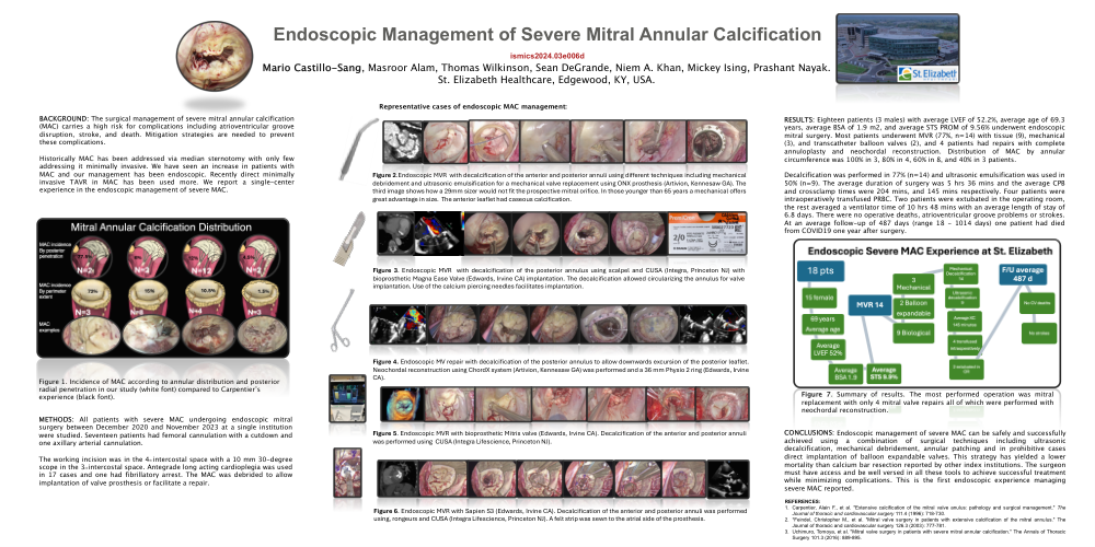
BACKGROUND: The surgical management of severe mitral annular calcification (MAC) carries a high risk for complications including atrioventricular groove disruption, stroke, and death. Mitigation strategies are needed to prevent these complications. Historically MAC has been addressed via median sternotomy with only few addressing it minimally invasive. We have seen an increase in patients with MAC and our management has been endoscopic. Recently direct minimally invasive TAVR in MAC has been used more. We report a single-center experience in the endoscopic management of severe MAC.
Figure 1. Incidence of MAC according to annular distribution and posterior radial penetration in our study (white font) compared to Carpentier’s experience (black font).
METHODS: All patients with severe MAC undergoing endoscopic mitral surgery between December 2020 and November 2023 at a single institution were studied. Seventeen patients had femoral cannulation with a cutdown and one axillary arterial cannulation. The working incision was in the 4th intercostal space with a 10 mm 30-degree scope in the 3rdintercostal space. Antegrade long acting cardioplegia was used in 17 cases and one had fibrillatory arrest. The MAC was debrided to allow implantation of valve prosthesis or facilitate a repair.
Figure 2.Endoscopic MVR with decalcification of the anterior and posterior annuli using different techniques including mechanical debridement and ultrasonic emulsification for a mechanical valve replacement using ONX prosthesis (Artivion, Kennesaw GA). The third image shows how a 29mm sizer would not fit the prospective mitral orifice. In those younger than 65 years a mechanical offers great advantage in size. The anterior leaflet had caseous calcification.
Figure 3. Endoscopic MVR with decalcification of the posterior annulus using scalpel and CUSA (Integra, Princeton NJ) with bioprosthetic Magna Ease Valve (Edwards, Irvine CA) implantation. The decalcification allowed circularizing the annulus for valve implantation. Use of the calcium piercing needles facilitates implantation.
Figure 4. Endoscopic MV repair with decalcification of the posterior annulus to allow downwards excursion of the posterior leaflet. Neochordal reconstruction using ChordX system (Artivion, Kennesaw GA) was performed and a 36 mm Physio 2 ring (Edwards, Irvine CA).
Figure 5. Endoscopic MVR with bioprosthetic Mitris valve (Edwards, Irvine CA). Decalcification of the anterior and posterior annuli was performed using CUSA (Integra Lifescience, Princeton NJ).
Figure 6. Endoscopic MVR with Sapien S3 (Edwards, Irvine CA). Decalcification of the anterior and posterior annuli was performed using, rongeurs and CUSA (Integra Lifescience, Princeton NJ). A felt strip was sewn to the atrial side of the prosthesis.
RESULTS: Eighteen patients (3 males) with average LVEF of 52.2%, average age of 69.3 years, average BSA of 1.9 m2, and average STS PROM of 9.56% underwent endoscopic mitral surgery. Most patients underwent MVR (77%, n=14) with tissue (9), mechanical (3), and transcatheter balloon valves (2), and 4 patients had repairs with complete annuloplasty and neochordal reconstruction. Distribution of MAC by annular circumference was 100% in 3, 80% in 4, 60% in 8, and 40% in 3 patients. Decalcification was performed in 77% (n=14) and ultrasonic emulsification was used in 50% (n=9). The average duration of surgery was 5 hrs 36 mins and the average CPB and crossclamp times were 204 mins, and 145 mins respectively. Four patients were intraoperatively transfused PRBC. Two patients were extubated in the operating room, the rest averaged a ventilator time of 10 hrs 48 mins with an average length of stay of 6.8 days. There were no operative deaths, atrioventricular groove problems or strokes. At an average follow-up of 487 days (range 18 - 1014 days) one patient had died from COVID19 one year after surgery.
Figure 7. Summary of results. The most performed operation was mitral replacement with only 4 mitral valve repairs all of which were performed with neochordal reconstruction.
CONCLUSIONS: Endoscopic management of severe MAC can be safely and successfully achieved using a combination of surgical techniques including ultrasonic decalcification, mechanical debridement, annular patching and in prohibitive cases direct implantation of balloon expandable valves. This strategy has yielded a lower mortality than calcium bar resection reported by other index institutions. The surgeon must have access and be well versed in all these tools to achieve successful treatment while minimizing complications. This is the first endoscopic experience managing severe MAC reported.
REFERENCES: