225 posters, 34 videos, 18 topics, 14 sessions, 251 authors, 55 institutions
ePostersLive by SciGen Technologies S.A. All rights reserved.
ISMICS 2024 Annual Meeting
29 May - 1 June, 2024 | Athens, Greece

D.44
Necrotizing Chest Wall Infection With Extension Into Breast Tissue Following Minimally Invasive Direct Coronary Artery Bypass Surgery
Poster Presenter
Authors
Affiliations
Part of Topic
Coronary Revascularization
Necrotizing chest wall infection with extension into breast tissue following minimally invasive direct coronary artery bypass surgery
Adin S.C. Reisner MD, Robin Willard MD, Nirav C. Patel MD
Lenox Hill Hospital, Northwell Health
Minimally invasive coronary artery bypass grafting is an effective way of addressing multi-vessel cardiac disease while avoiding a sternotomy, with rates of post operative infection as low as 0.3% [1]. However, patients with uncontrolled diabetes are at elevated risk of developing necrotizing infections [2] and the impact, if any, when combined with relative tissue ischemia after a left internal mammary artery harvest is unknown.
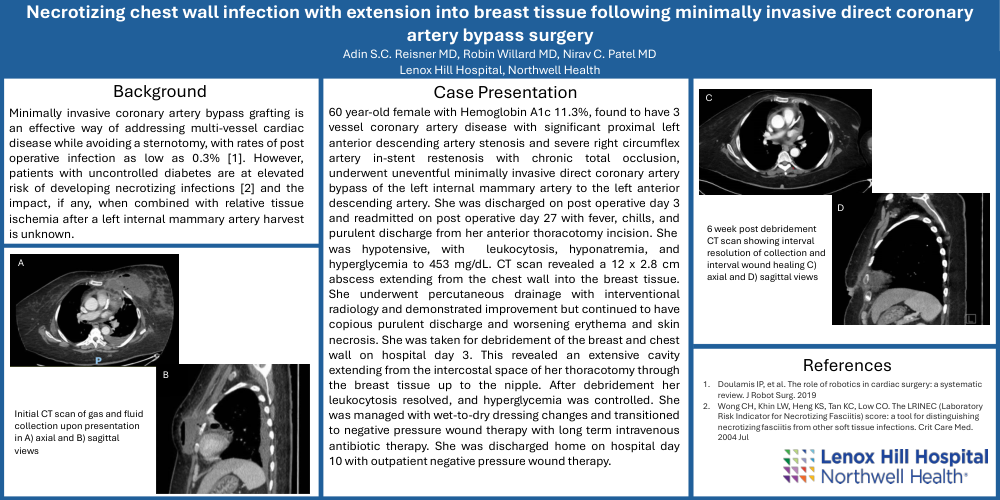
60 year-old female with Hemoglobin A1c 11.3%, found to have 3 vessel coronary artery disease with significant proximal left anterior descending artery stenosis and severe right circumflex artery in-stent restenosis with chronic total occlusion, underwent uneventful minimally invasive direct coronary artery bypass of the left internal mammary artery to the left anterior descending artery. She was discharged on post operative day 3 and readmitted on post operative day 27 with fever, chills, and purulent discharge from her anterior thoracotomy incision. She was hypotensive, with leukocytosis, hyponatremia, and hyperglycemia to 453 mg/dL. CT scan revealed a 12 x 2.8 cm abscess extending from the chest wall into the breast tissue. She underwent percutaneous drainage with interventional radiology and demonstrated improvement but continued to have copious purulent discharge and worsening erythema and skin necrosis. She was taken for debridement of the breast and chest wall on hospital day 3. This revealed an extensive cavity extending from the intercostal space of her thoracotomy through the breast tissue up to the nipple. After debridement her leukocytosis resolved, and hyperglycemia was controlled. She was managed with wet-to-dry dressing changes and transitioned to negative pressure wound therapy with long term intravenous antibiotic therapy. She was discharged home on hospital day 10 with outpatient negative pressure wound therapy.