225 posters, 34 videos, 18 topics, 14 sessions, 251 authors, 55 institutions
ePostersLive by SciGen Technologies S.A. All rights reserved.
ISMICS 2024 Annual Meeting
29 May - 1 June, 2024 | Athens, Greece

D.33
Surgical Outcome Of Post-infarction Left Ventricular Free Wall Rupture
Poster Presenter
Authors
Part of Topic
Coronary Revascularization
Surgical outcome of post-infarction left ventricular free wall rupture
Chan-Young Na M.D.
Hallym University, Korea
Introduction
Cardiac rupture after acute myocardial infarction, which includes ventricular free wall rupture, septal rupture and papillary muscle rupture, could result in lethal complications.
Early recognition and aggressive treatment are recommended. However, its clinical results outcomes are poorly studied because of the condition’s rarity. Here author review the surgical results of ischemic free wall rupture of the left ventricle.
Patient Profile
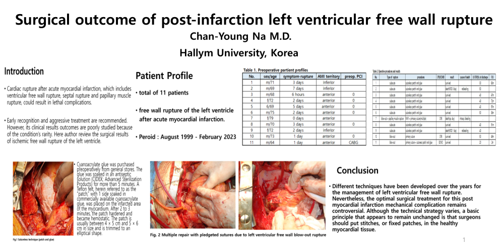
- total of 11 patients
- free wall rupture of the left ventricle after acute myocardial infarction.
- Peroid : August 1999 - February 2023
Method
Cyanoacrylate glue was purchased preoperatively from general stores. The glue was soaked in an antiseptic solution (CIDEX; Advanced Sterilization Products) for more than 5 minutes. A Teflon felt, herein referred to as the “patch,” with 1 side soaked in commercially available cyanoacrylate glue, was placed on the infarcted area of the myocardium. After 2 to 3 minutes, the patch hardened and became hemostatic. The patch is usually between 4 × 5 cm and 5 × 6 cm in size and is trimmed to an elliptical shape.
Conclusion
Different techniques have been developed over the years for the management of left ventricular free wall rupture. Nevertheless, the optimal surgical treatment for this post myocardial infarction mechanical complication remains controversial. Although the technical strategy varies, a basic principle that appears to remain unchanged is that surgeons should put stitches, or fixed patches, in the healthy myocardial tissue.