167 posters, 28 videos, 9 topics, 12 sessions, 337 authors, 160 institutions
ePostersLive by SciGen Technologies S.A. All rights reserved.
ISMICS 2023 Annual Meeting
31 May - 3 June, 2023 | Boston, MA

PC89
Minimally Invasive Surgical Valve-in-Ring
Poster Presenter
Authors
Affiliations
Part of Topic
Valve - Surgical Valves
Minimally Invasive Surgical Valve-in-Ring
Background
Although several causes lead to reoperation in patients with previously implanted cardiac bioprosthesis, the most common cause is structural failure. The removal of an aortic and/or a mitral prosthesis can be a delicate surgery because patients may have a deteriorating or fragile clinical condition (1,2). Prosthesis in general, have improved in design, durability, and function; as technology and comprehensive knowledge of valvular function advances, so does the quality and surgical technique employed not only in replacement but also in repair (3).
Patients with previous mitral valve surgery tend to be older and sicker than those having a primary valve replacement. In addition to the increased risk of reoperation, mitral valve surgery is associated with cardiac rupture at the atrioventricular junction or posterior ventricular wall where a strut could potentially be embedded, or damage to the circumflex artery during explantation maneuvers of a prosthesis. Furthermore, the remaining annulus can be very weak, and perivalvular leaks may ensue, increasing morbidity and mortality (1,2).
Geha et al. described in 1991 (4) the surgical technique for replacement without explantation of a degenerated mitral bioprosthesis, to reduce the hazards associated with the complete removal by leaving the stent and support in place(4). Previous descriptions of surgical implantation of a mechanical prosthesis sutured to an old bioprosthesis ring were also described by Stassano et al. however, this superimposed position only works when the new valve is one or two sizes smaller (2).
As previously described, the major risks of reoperation in the mitral position are the removal of prosthetic materials, especially when there is calcification of the tissue ingrowth, strut incorporation to the myocardial wall, or valve-to-annulus mismatch (4). Although these complications are described in a valve degeneration scenario, they can also happen in patients with another type of foreign material, as in the described case with an annuloplasty ring.
Results
The postoperative course was uneventful, the patient was extubated in the ICU on postop day 1, and physical and pulmonary rehabilitation was initiated as soon as residual sedation wore off. Thoracic drains were taken off on day 2 with no significant postoperative bleeding.
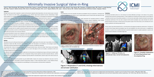
He developed pneumonia with increased oxygen supply requirements but responded positively to antibiotic therapy and was discharged on postoperative day 10. He continues physical rehabilitation, which includes pulmonary and swallowing therapy; follow-up echo shows acceptable hemodynamic performance
Conclusions.
As previously described by Geha et al., this technique has technical difficulties and hinges on the absence of any previous paravalvular leak around the remaining prosthesis, so when employed, this should be the foremost aspect to ensure (Figure 3) ; secondly, the entire tissue of the previous prosthesis, or in this case the native leaflet, should be removed to ensure proper hemodynamic performance of the new valve. Leaving the cloth-covered annuloplasty ring in place eliminates the need for extensive dissection and results in minimal trauma to the posterior aspect of the mitral annulus, and so eliminates the risk of surgical complications and reducing surgical time (5).
Previous reports have demonstrated that this is a safe alternative with adequate hemodynamic performances. The fixation of one valve to other foreign materials (annuloplasty ring in this case) embedded in the native mitral annulus is a straightforward procedure; it is proven to be an excellent alternative to avoid the dreaded hazards of complete explantation in patients with extensive annular calcification or the elderly-fragile state.
1.Geha AS, Massad MG, Snow NJ. Replacement of Degenerated Mitral and Aortic BioprosthesesWithout Explantation. 2001.
2.Stassano P, Losi MA, Golino A, Gagliardi C, Iorio D, Marzullo M, et al. Bioprosthesis replacement with mechanical valve implantation on the bioprosthetic ring Surgical and 2D echo considerations. Vol. 7. Springer-Verlag; 1993.
3.Miyairi S, Koide M, Kunii Y, Tsuda K, Ohashi Y, Harada T. Redo mitral valve replacement using the valve-on-valve method. Asian Cardiovasc ThoracAnn. 2015 Jul 19;23(6):707–9.
4.GEHA A, LEE J. New approach for replacement of degenerated mitral bioprostheses. European Journal of Cardio-Thoracic Surgery. 1996;10(12):1090–1063.
5.Geha AS, Massad MG, Snow NJ. Replacement of degenerated mitral and aortic bioprostheseswithout explantation. Ann Thorac Surg. 2001 Nov;72(5):1509–14.