167 posters, 28 videos, 9 topics, 12 sessions, 337 authors, 160 institutions
ePostersLive by SciGen Technologies S.A. All rights reserved.
ISMICS 2023 Annual Meeting
31 May - 3 June, 2023 | Boston, MA

PC43
Robotic Transmitral Septal Myectomy And Secondary Chord Resection For Hypertrophic Obstructive Cardiomyopathy
Poster Presenter
Authors
Affiliations
Part of Topic
Valve - Surgical Valves
Video
Robotic Transmitral Septal Myectomy And Secondary Chord Resection For Hypertrophic Obstructive Cardiomyopathy
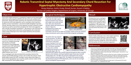
Trans-aortic septal myectomy is a standard of surgical care for hypertrophic obstructive cardiomyopathy (HOCM) with left ventricular outflow (LVOT) obstruction. Along with septal myectomy, the importance of intervention for abnormal mitral valve apparatus such as secondary chord resection1, papillary muscle reorientation2, and anterior leaflet elongation3 has been noticed. Recently, robotic trans-mitral approach has been utilized for septal myectomy as well as concomitant procedures for mitral valve apparatus with a benefit of excellent visualization4,5. We present a case of 59-year-old woman with hypertrophic obstructive cardiomyopathy who underwent robotic totally endoscopic trans-mitral septal myectomy and secondary chord resection.
A 59-year-old woman with HOCM came to our hospital presenting with shortness of breath. Transthoracic echocardiography revealed severe LVOT obstruction with maximum gradient of 83 mmHg and severe mitral regurgitation. Interventricular septal thickness was 1.6 cm. Trans-esophageal echo demonstrated thick secondary chords of the anterior mitral leaflet playing a primary role to tether the anterior mitral leaflet (AML) toward the LVOT resulting in severe obstruction (Figure 1). Severe mitral regurgitation was caused by systolic anterior motion of the AML and posterior mitral leaflet prolapse
The patient was placed supine with a roll under the right scapula. General anesthesia was induced with single lumen endo-tracheal tube. Ports were placed (Our standard robotic port placement is shown in Figure 2). Cardiopulmonary bypass was established with femoral cannulation. The Da Vinci Xi robot was docked. Cardiac arrest was achieved using a transthoracic aortic clamp and Del Nido antegrade cardioplegia. The mitral valve was exposed via left atriotomy. The AML was detached from commissure to commissure using sharp scissors, leaving a 3-mm cuff attached to the annulus (Figure 3A). The fibrotic secondary chordae were carefully identified and excised as much as possible (figure 3B). The interventricular septum was carefully exposed through this plane using the dynamic atrial retractor, and hypertrophied septal muscle was resected sharply (Figure 3C). The AML was primarily reattached to the annulus using 5-0 prolene suture (Figure 3D). The height of the posterior leaflet was shortened using CV-4 PTFE NeoChords, and a size 32-mm semirigid partial annuloplasty ring was placed.
Postoperative trans-esophageal echocardiography showed max gradient across the LVOT was 8 mmHg and no significant mitral regurgitation. The patient was discharged on postoperative day 2 and returned to full activity in 14 days.
Robotic totally endoscopic trans-mitral septal myectomy and secondary chord resection is safe and feasible procedure for HOCM with LVOT obstruction. Robotic approach would be suitable for mitral valve exposure and intervention of mitral subvalvular apparatus.