167 posters, 28 videos, 9 topics, 12 sessions, 337 authors, 160 institutions
ePostersLive by SciGen Technologies S.A. All rights reserved.
ISMICS 2023 Annual Meeting
31 May - 3 June, 2023 | Boston, MA

D26
Why Does A 14-year-old Girl Have Pericardial Effusion
Poster Presenter
Part of Topic
Pediatric - Cardiac, Cardiovascular & Thoracic
Why does a 14-year-old girl have pericardial effusion
Huanlei Huang, Lishan Zhong, Weidong Lin, Hongwen Fei
Guangdong Cardiovascular Institution, Guangdong Provincial People’s Hospital, Guangdong Academy of Medical Sciences
Objective
Post-cardiac injury syndromes (PCIS), which carry a higher risk of consequences, can be quite complex. The most terrifying PCIS consequence is ventricular perforation (VP). If it does occur, prompt treatment can be facilitated by being aware of the potential impact and paying close attention to patient monitoring. We presented a special case that included a myocardial contrast echocardiogram (MCE)-described blood pericardial effusion related to VP and the successful closure of a right ventricular pseudoaneurysm.
Results
To accurately diagnose pericardial effusion with an unknown origin, several diagnostic techniques must be employed. Although heart rupture linked to cardiac damage syndrome is extremely uncommon, we should exercise caution. Emergency thoracotomies are advised for situations of life-threatening heart and chest penetrating trauma rather than pericardiocentesis and drainage as the first line of treatment to increase survival.
Methods
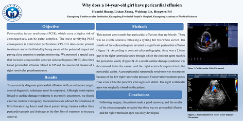
This patient consistently has pericardial effusions that are bloody. There was no visible soreness following a cycling fall two weeks earlier. The results of the echocardiogram revealed a significant pericardial effusion (Figure 1). According to contrast echocardiography, there was a 2.6mm gap in the right ventricular apex through which the contrast agent reached the pericardial cavity (Figure 2). As a result, cardiac damage syndrome was determined to be the cause, and the right ventricle ruptured into the pericardial cavity. Acute pericardial tamponade syndrome was not present because of the low right ventricular pressure. Conservative treatment poses risks even while the patient's vital signs are stable. The right ventricular apex was surgically closed on the patient.
Conclusions
Following surgery, the patient made a good recovery, and the results of the ultrasonography revealed that there was no pericardial effusion and the right ventricular apex was fully developed.