477 posters, 14 topics, 2,065 authors, 1,055 institutions
ePostersLive by SciGen Technologies S.A. All rights reserved.
The Joint EBJIS-MSIS 2026 Conference
17 - 19 September, 2026 | Porto, Portugal

EP047
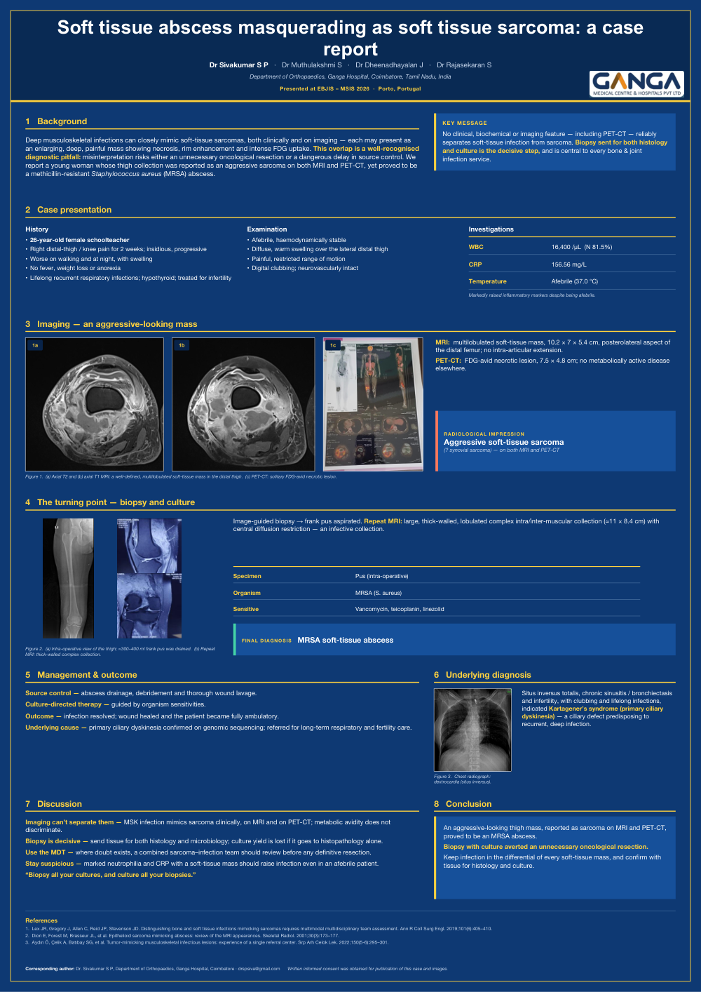
Soft tissue abscess masquerading as Soft tissue sarcoma : A case report
Poster Presenter
Authors
Affiliations
Part of Topic
Case Report
Soft tissue abscess masquerading as soft tissue sarcoma: a case report
Dr Sivakumar S P · Dr Muthulakshmi S · Dr Dheenadhayalan J · Dr Rajasekaran S
Department of Orthopaedics, Ganga Hospital, Coimbatore, Tamil Nadu, India
Corresponding author: Dr Sivakumar S P · drspsiva@gmail.com
Keywords
Soft-tissue abscess; soft-tissue sarcoma; tumour mimic; musculoskeletal infection; MRSA; Staphylococcus aureus; MRI; PET-CT; diagnostic pitfall; image-guided biopsy and culture; source control; primary ciliary dyskinesia; Kartagener syndrome; bone and joint infection.
Abstract
- Background — Deep musculoskeletal infection can closely mimic soft-tissue sarcoma, both clinically and on imaging; misinterpretation risks either an unnecessary oncological resection or a dangerous delay in source control.
- Case — A 26-year-old woman presented with two weeks of insidious, progressive right distal-thigh / knee pain and swelling, worse at night. She was afebrile with digital clubbing; WBC was 16,400/µL (81.5% neutrophils) and CRP 156.56 mg/L.
- Imaging — MRI showed a multilobulated soft-tissue mass (10.2 × 7 × 5.4 cm) of the posterolateral distal femur, and PET-CT an FDG-avid necrotic lesion (7.5 × 4.8 cm) — both reported as an aggressive soft-tissue sarcoma (? synovial).
- Turning point — Image-guided biopsy yielded frank pus; culture grew MRSA. Repeat MRI showed a thick-walled complex collection (≈11 × 8.4 cm).
- Management & outcome — Drainage, debridement and lavage with culture-directed antibiotics; the infection resolved and the patient became ambulatory. Underlying primary ciliary dyskinesia (Kartagener's syndrome) was confirmed on genomic sequencing.
- Conclusion — No clinical, biochemical or imaging feature — including PET-CT — reliably separates infection from sarcoma. Biopsy sent for both histology and culture is decisive.
Background
Deep musculoskeletal infections can closely mimic soft-tissue sarcomas, both clinically and on imaging — each may present as an enlarging, deep, painful mass showing necrosis, rim enhancement and intense FDG uptake. This overlap is a well-recognised diagnostic pitfall: misinterpretation risks either an unnecessary oncological resection or a dangerous delay in source control. We report a young woman whose thigh collection was reported as an aggressive sarcoma on both MRI and PET-CT, yet proved to be a methicillin-resistant Staphylococcus aureus (MRSA) abscess.
Case presentation
History
- 26-year-old female schoolteacher.
- Right distal-thigh / knee pain for 2 weeks; insidious, progressive.
- Worse on walking and at night, with swelling.
- No fever, weight loss or anorexia.
- Lifelong recurrent respiratory infections; hypothyroid; treated for infertility.
Examination
- Afebrile, haemodynamically stable.
- Diffuse, warm swelling over the lateral distal thigh.
- Painful, restricted range of motion.
- Digital clubbing; neurovascularly intact.
Investigations
WBC: 16,400 /µL (neutrophils 81.5%)
CRP: 156.56 mg/L
Temperature: Afebrile (37.0 °C)
Markedly raised inflammatory markers despite being afebrile.
Imaging — an aggressive-looking mass
- MRI: multilobulated soft-tissue mass, 10.2 × 7 × 5.4 cm, posterolateral aspect of the distal femur; no intra-articular extension.
- PET-CT: FDG-avid necrotic lesion, 7.5 × 4.8 cm; no metabolically active disease elsewhere.
Radiological impression: aggressive soft-tissue sarcoma (? synovial sarcoma) — on both MRI and PET-CT.
The turning point — biopsy and culture
Image-guided biopsy yielded frank pus. Repeat MRI showed a large, thick-walled, lobulated complex intra/inter-muscular collection (≈11 × 8.4 cm) with central diffusion restriction — an infective collection.
Specimen: Pus (intra-operative)
Organism: MRSA (Staphylococcus aureus)
Sensitive: Vancomycin, teicoplanin, linezolid
Final diagnosis: MRSA soft-tissue abscess.
Management & outcome
- Source control — abscess drainage, debridement and thorough wound lavage.
- Culture-directed therapy — guided by organism sensitivities.
- Outcome — infection resolved; wound healed and the patient became fully ambulatory.
- Underlying cause — primary ciliary dyskinesia confirmed on genomic sequencing; referred for long-term respiratory and fertility care.
Underlying diagnosis
Situs inversus totalis, chronic sinusitis / bronchiectasis and infertility, with clubbing and lifelong infections, indicated Kartagener's syndrome (primary ciliary dyskinesia) — a ciliary defect predisposing to recurrent, deep infection.
Discussion — relevance to musculoskeletal infection practice
- Imaging can't separate them — musculoskeletal infection mimics sarcoma clinically, on MRI and on PET-CT; metabolic avidity does not discriminate.
- Biopsy is decisive — send tissue for both histology and microbiology; culture yield is lost if it goes to histopathology alone.
- Use the MDT — where doubt exists, a combined sarcoma–infection team should review before any definitive resection.
- Stay suspicious — marked neutrophilia and CRP with a soft-tissue mass should raise infection even in an afebrile patient.
“Biopsy all your cultures, and culture all your biopsies.”
Key message
No clinical, biochemical or imaging feature — including PET-CT — reliably separates soft-tissue infection from sarcoma. Biopsy sent for both histology and culture is the decisive step, and is central to every bone & joint infection service.
Conclusion
An aggressive-looking thigh mass, reported as sarcoma on MRI and PET-CT, proved to be an MRSA abscess. Biopsy with culture averted an unnecessary oncological resection. Keep infection in the differential of every soft-tissue mass, and confirm with tissue for histology and culture.
Figure legends
Figure 1. (a) Axial T2 and (b) axial T1 MRI: a well-defined, multilobulated soft-tissue mass in the distal thigh. (c) PET-CT: solitary FDG-avid necrotic lesion.
Figure 2. (a) Intra-operative view of the thigh; ≈300–400 ml frank pus was drained. (b) Repeat MRI: thick-walled complex collection.
Figure 3. Chest radiograph: dextrocardia (situs inversus).
References
1. Lex JR, Gregory J, Allen C, Reid JP, Stevenson JD. Distinguishing bone and soft tissue infections mimicking sarcomas requires multimodal multidisciplinary team assessment. Ann R Coll Surg Engl. 2019;101(6):405–410.
2. Dion E, Forest M, Brasseur JL, et al. Epithelioid sarcoma mimicking abscess: review of the MRI appearances. Skeletal Radiol. 2001;30(3):173–177.
3. Aydın Ö, Çelik A, Batıbay SG, et al. Tumor-mimicking musculoskeletal infectious lesions: experience of a single referral center. Srp Arh Celok Lek. 2022;150(5-6):295–301.
Written informed consent was obtained for publication of this case and images.