477 posters, 14 topics, 2,065 authors, 1,055 institutions
ePostersLive by SciGen Technologies S.A. All rights reserved.
The Joint EBJIS-MSIS 2026 Conference
17 - 19 September, 2026 | Porto, Portugal

EP481
An Unexpected Pancoast Mimic: A Septic Costovertebral Joint
Poster Presenter
Part of Topic
Spinal Infections
Septic arthritis of the costovertebral joint (CVJ) is a rare and underrecognized entity that may mimic apical lung malignancy, creating a diagnostic challenge. We present a 69-year-old man with diabetes, hepatitis C, and prior anterior cervical spine fusion who developed acute confusion and right shoulder pain. Blood cultures grew methicillin-sensitive Staphylococcus aureus (MSSA), which cleared within 24 hours of antibiotic therapy. Despite treatment, persistent shoulder pain radiating to the neck and sternoclavicular region prompted imaging.
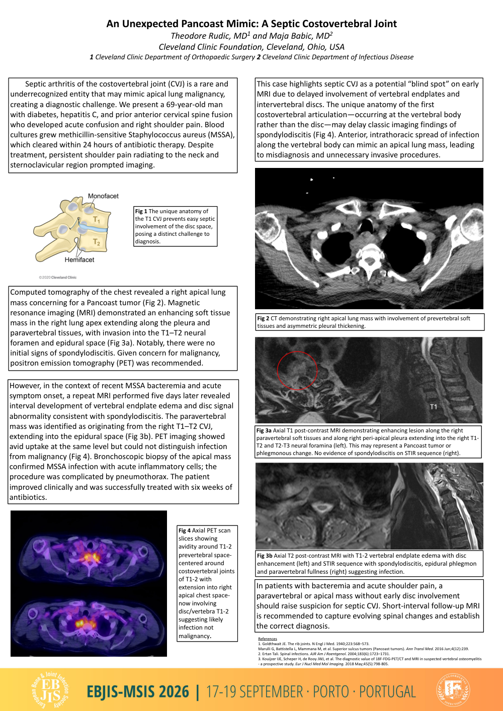
Computed tomography of the chest revealed a right apical lung mass concerning for a Pancoast tumor (Fig 2). Magnetic resonance imaging (MRI) demonstrated an enhancing soft tissue mass in the right lung apex extending along the pleura and paravertebral tissues, with invasion into the T1–T2 neural foramen and epidural space (Fig 3a). Notably, there were no initial signs of spondylodiscitis. Given concern for malignancy, positron emission tomography (PET) was recommended.
However, in the context of recent MSSA bacteremia and acute symptom onset, a repeat MRI performed five days later revealed interval development of vertebral endplate edema and disc signal abnormality consistent with spondylodiscitis. The paravertebral mass was identified as originating from the right T1–T2 CVJ, extending into the epidural space (Fig 3b). PET imaging showed avid uptake at the same level but could not distinguish infection from malignancy (Fig 4). Bronchoscopic biopsy of the apical mass confirmed MSSA infection with acute inflammatory cells; the procedure was complicated by pneumothorax. The patient improved clinically and was successfully treated with six weeks of antibiotics.
This case highlights septic CVJ as a potential “blind spot” on early MRI due to delayed involvement of vertebral endplates and intervertebral discs. The unique anatomy of the first costovertebral articulation—occurring at the vertebral body rather than the disc—may delay classic imaging findings of spondylodiscitis (Fig 4). Anterior, intrathoracic spread of infection along the vertebral body can mimic an apical lung mass, leading to misdiagnosis and unnecessary invasive procedures.
In patients with bacteremia and acute shoulder pain, a paravertebral or apical mass without early disc involvement should raise suspicion for septic CVJ. Short-interval follow-up MRI is recommended to capture evolving spinal changes and establish the correct diagnosis.