477 posters, 14 topics, 2,065 authors, 1,055 institutions
ePostersLive by SciGen Technologies S.A. All rights reserved.
The Joint EBJIS-MSIS 2026 Conference
17 - 19 September, 2026 | Porto, Portugal

EP127
Actinomycotic mycetoma (Madura foot) mimicking a soft tissue tumor after minor trauma: a diagnostic challenge
Poster Presenter
Authors
Jawhar Ben Dhief, Youssef Othman, Azer Ben Salah, Adnene Toumi, Saber Rabhi, Makrem Zrig, abderrazak abid
Affiliations
Infectious Diseases Department, Fattouma Bourguiba University Hospital, Tunisia, The Orthopedics and Traumatology Department, Habib Bourguiba University Hospital, Sfax, The Orthopedics and Traumatology Department, Fattouma Bourguiba University Hospital, Tunisia, parasitology laboratory, Fattouma Bourguiba University Hospital, Tunisia
Part of Topic
Fracture-Related Infections
Introduction : Mycetoma, commonly known as Madura foot, is a chronic granulomatous infection endemic in tropical and subtropical regions. The disease is caused by either aerobic bacteria (actinomycetoma, ~60% of cases) or fungi (eumycetoma). Although primarily affecting rural and agricultural populations, mycetoma remains underrecognized in clinical practice. Its indolent clinical course, nonspecific presentation, and tumor-like appearance frequently lead to diagnostic delays and erroneous interpretation as neoplastic or nonspecific inflammatory lesions. Accurate diagnosis requires high clinical suspicion, appropriate imaging, and microbiological confirmation.
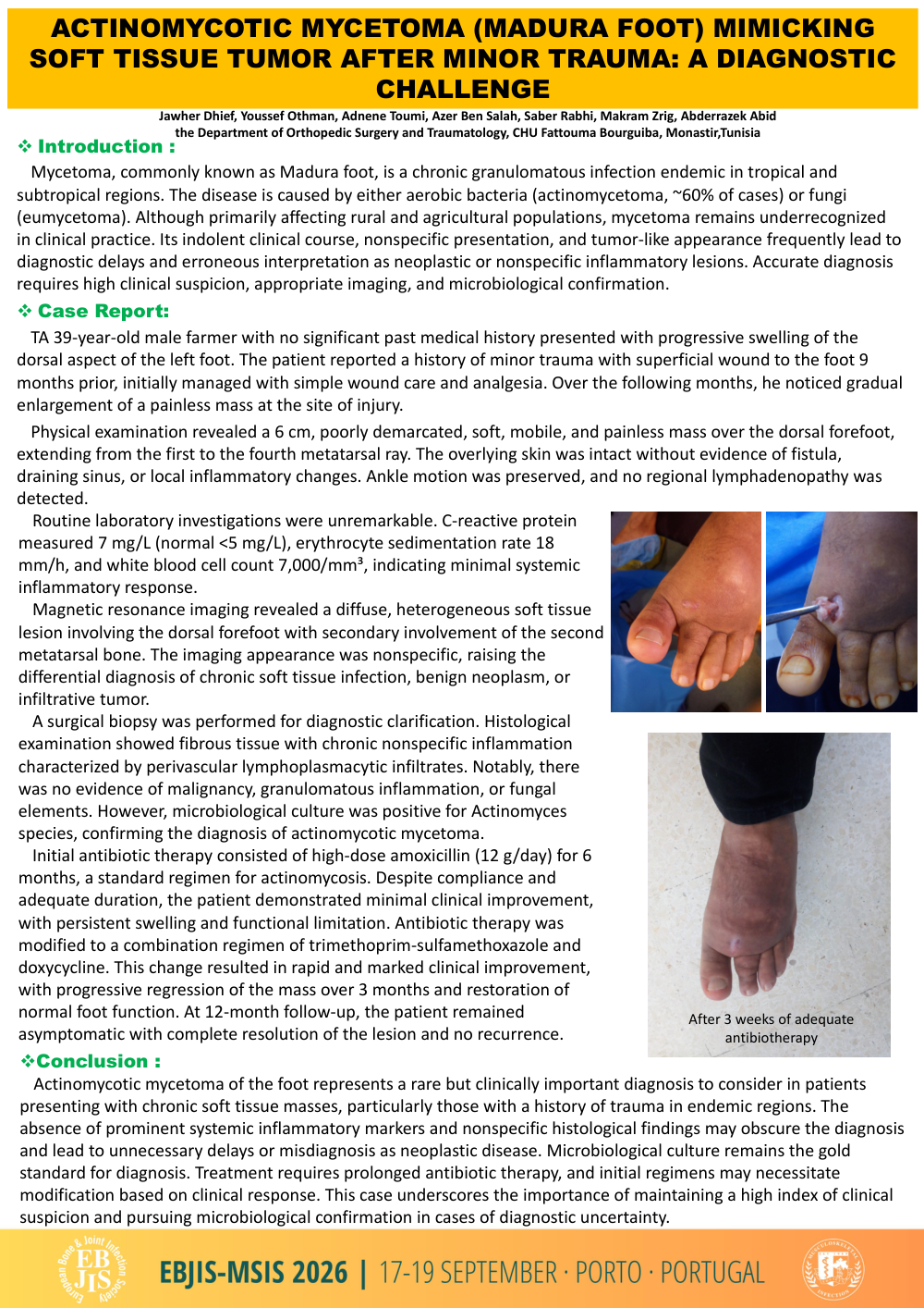
Case Report: TA 39-year-old male farmer with no significant past medical history presented with progressive swelling of the dorsal aspect of the left foot. The patient reported a history of minor trauma with superficial wound to the foot 9 months prior, initially managed with simple wound care and analgesia. Over the following months, he noticed gradual enlargement of a painless mass at the site of injury. Physical examination revealed a 6 cm, poorly demarcated, soft, mobile, and painless mass over the dorsal forefoot, extending from the first to the fourth metatarsal ray. The overlying skin was intact without evidence of fistula, draining sinus, or local inflammatory changes. Ankle motion was preserved, and no regional lymphadenopathy was detected. Routine laboratory investigations were unremarkable. C-reactive protein measured 7 mg/L (normal <5 mg/L), erythrocyte sedimentation rate 18 mm/h, and white blood cell count 7,000/mm³, indicating minimal systemic inflammatory response. Magnetic resonance imaging revealed a diffuse, heterogeneous soft tissue lesion involving the dorsal forefoot with secondary involvement of the second metatarsal bone. The imaging appearance was nonspecific, raising the differential diagnosis of chronic soft tissue infection, benign neoplasm, or infiltrative tumor. A surgical biopsy was performed for diagnostic clarification. Histological examination showed fibrous tissue with chronic nonspecific inflammation characterized by perivascular lymphoplasmacytic infiltrates. Notably, there was no evidence of malignancy, granulomatous inflammation, or fungal elements. However, microbiological culture was positive for Actinomyces species, confirming the diagnosis of actinomycotic mycetoma. Initial antibiotic therapy consisted of high-dose amoxicillin (12 g/day) for 6 months, a standard regimen for actinomycosis. Despite compliance and adequate duration, the patient demonstrated minimal clinical improvement, with persistent swelling and functional limitation. Antibiotic therapy was modified to a combination regimen of trimethoprim-sulfamethoxazole and doxycycline. This change resulted in rapid and marked clinical improvement, with progressive regression of the mass over 3 months and restoration of normal foot function. At 12-month follow-up, the patient remained asymptomatic with complete resolution of the lesion and no recurrence.
Conclusion : Actinomycotic mycetoma of the foot represents a rare but clinically important diagnosis to consider in patients presenting with chronic soft tissue masses, particularly those with a history of trauma in endemic regions. The absence of prominent systemic inflammatory markers and nonspecific histological findings may obscure the diagnosis and lead to unnecessary delays or misdiagnosis as neoplastic disease. Microbiological culture remains the gold standard for diagnosis. Treatment requires prolonged antibiotic therapy, and initial regimens may necessitate modification based on clinical response. This case underscores the importance of maintaining a high index of clinical suspicion and pursuing microbiological confirmation in cases of diagnostic uncertainty.