477 posters, 14 topics, 2,065 authors, 1,055 institutions
ePostersLive by SciGen Technologies S.A. All rights reserved.
The Joint EBJIS-MSIS 2026 Conference
17 - 19 September, 2026 | Porto, Portugal

EP101
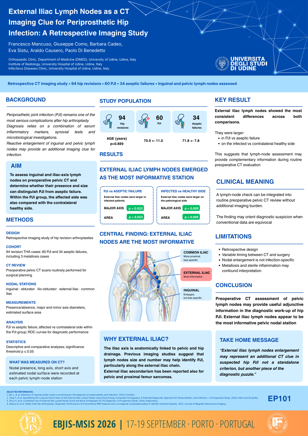
External Iliac Lymph Nodes as a CT Imaging Clue for Periprosthetic Hip Infection: A Retrospective Imaging Study
Poster Presenter
Authors
Francesco Mancuso, Giuseppe Como, Barbara Cadeo, Eva Sistu, Araldo Causero, Paolo Di Benedetto
Affiliations
Orthopedics Clinic, University Hospital, Udine, Italy; Medicine Department - DMED, University of Udine, Udine, Italy, Institute of Radiology, University Hospital, Udine, Italy; Medicine Department - DMED, University of Udine, Udine, Italy, Infectious Diseases Clinic, University Hospital, Udine, Italy, Orthopedics Clinic, DMED, University of Udine
Part of Topic
Diagnosis
Aim
Periprosthetic joint infection (PJI) is one of the most serious complications after hip arthroplasty. Alongside serum inflammatory markers, synovial tests, and microbiological investigations, imaging findings may provide additional diagnostic support. Enlargement of inguinal and pelvic lymph nodes, particularly along the external iliac axis, has recently been proposed as a possible indicator of infection. The aim of this study was to evaluate the role of inguinal and iliac-axis lymph nodes on preoperative pelvic CT in distinguishing PJI from aseptic failure and in supporting treatment follow-up.
Methods
This retrospective study included 94 hip revision arthroplasties: 60 for PJI and 34 for aseptic failure (including 3 metallosis). Preoperative pelvic CT scans, routinely performed for surgical planning, were reviewed to assess the presence and size of lymph nodes in the inguinal and iliac stations, including external iliac nodes. Measurements included major and minor axis diameters and estimated surface area. Lymph node findings were compared between infected and non-infected patients and, within the PJI group, between the affected and contralateral healthy side. Descriptive and comparative statistical analyses were performed, and receiver operating characteristic (ROC) curves were used to evaluate diagnostic performance.
Results
Among all nodal stations, although inguinal lymph nodes were significantly enlarged on the infected side compared to the contralateral healthy side, they did not clearly discriminate PJI from aseptic failure; in this respect, external iliac lymph showed the most consistent differences. In the comparison between PJI and aseptic failure, external iliac nodes were significantly larger in infected patients, both for major axis (p = 0.023) and area (p = 0.023).
Within the PJI group, comparison between the infected side and the contralateral healthy side confirmed significantly greater external iliac node size on the pathological side, again for both major axis (p = 0.005) and area (p = 0.005). Metallosis, as a cause of aseptic failure, may represent a confounding factor, although the cases in the present study are extremely limited.
Conclusions
Preoperative CT assessment of lymph nodes may provide useful adjunctive information in the diagnostic work-up of PJI. In particular, external iliac lymph nodes appear to be the most informative pelvic nodal station, showing reproducible dimensional differences in infected patients. Although not a standalone diagnostic criterion, their evaluation may represent an additional imaging clue in suspected periprosthetic hip infection.