99 posters, 2 videos, 5 audios, 13 topics, 7 sessions, 416 authors, 181 institutions
ePostersLive by SciGen Technologies S.A. All rights reserved.
The 21st Meeting of the Diabetic Foot Study Group 2026
24-26 September, 2026 | Florence, Italy

EP14
Radiological Detection of Advanced Gout in Patients with Diabetes and Foot Ulcers
Poster Presenter
Authors
Claus Rasmussen, Anne Marie Kelterberg-Wesenberg, Kasper Kofod Pedersen, Anne Grethe Jurik
Affiliations
North Denmark Regional Hospital, Rheumatology, Hj?rring, Denmark, North Denmark Regional Hospital, Centre for Clinical Research, , Hj?rring, Denmark, Clinical Department, Aalborg University, Aalborg, Denmark, North Denmark Regional Hospital, Radiology, Hj?rring, Denmark, North Denmark Regional Hospital, Centre for Clinical Research, , Hj?rring, Denmark
Part of Topic
Foot clinics
Radiological Detection of Advanced Gout in Patients with Diabetes and Foot Ulcers
Background:
Gout is often overlooked in patients with diabetes and foot ulcers, as neuropathy and ischemia can mask its typical inflammatory presentation. Undertreated gout frequently leads to tophus formation in the feet, which may ulcerate. Gout independently increases the risk of lower-extremity amputations by 50%. Urate-lowering therapy can prevent and dissolve tophi, making early diagnosis crucial. While conventional radiography (CR) and dual-energy CT (DECT) can detect advanced gout with osseous changes, their diagnostic utility in this population remains unevaluated.
Methods:
This study examined patients with diabetes referred to a tertiary foot-ulcer clinic. Patients underwent foot assessment with CR, supplemented by DECT when available. Radiological findings were assessed blind to clinical gout status and classified as consistent/inconsistent with gout. Clinical gout (urate-lowering therapy + clinical assessment) was the reference standard.
Results:
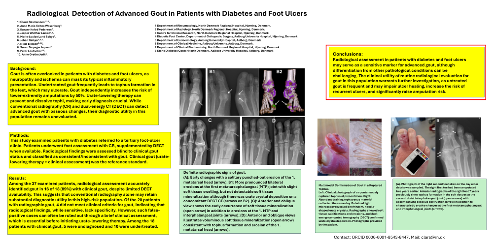
Among the 37 examined patients, radiological assessment accurately identified gout in 16 of 18 (89%) with clinical gout, despite limited DECT availability. This suggests that conventional radiography alone may retain substantial diagnostic utility in this high-risk population. Of the 20 patients with radiographic gout, 4 did not meet clinical criteria for gout, indicating that radiological findings, while sensitive, lack specificity. However, such false-positive cases can often be ruled out through a brief clinical assessment, which is essential before initiating urate-lowering therapy. Among the 18 patients with clinical gout, 5 were undiagnosed and 10 were undertreated.
Conclusions:
Radiological assessment in patients with diabetes and foot ulcers may serve as a sensitive marker for advanced gout, although differentiation from other pathological conditions can be challenging. The clinical utility of routine radiological evaluation for gout in this population warrants further investigation, as untreated gout is frequent and may impair ulcer healing, increase the risk of recurrent ulcers, and significantly raise amputation risk.