20 posters, 13 topics, 1 sessions, 54 authors, 27 institutions
ePostersLive by SciGen Technologies S.A. All rights reserved.
Clinical Radiology Audit and Quality Improvement ePoster Competitions 2026
30 September, 2026 | London, UK

79
The diagnostic accuracy of pelvic and hip radiographs in early detection of femoroacetabular impingement using MRI hip as standard investigation for patients presenting with painful hip.
Poster Presenter
Authors
Affiliations
Part of Topic
MSK
Title:
The diagnostic accuracy of pelvic and hip radiographs in early detection of femoroacetabular impingement using MRI hip as standard investigation for patients presenting with painful hip
Authors and Affiliation: (Amun Iqbal, Hafiza Mehr un Nisa) Dr. Sulaiman Alhabib HospitalRiyadh, Saudi Arabia
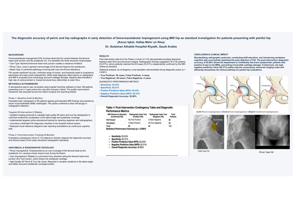
Background: Femoroacetabular impingement (FAI) involves abnormal hip contact between the femoral head-neck and acetabular rim, classified into Cam, Pincer, or Mixed types. It causes hip pain and restricted range of motion, often leading to labral tears and early osteoarthritis. Initial diagnosis relies on radiographs and MRI to evaluate bone morphology and joint cartilage damage. This quality improvement (QI) project aimed to optimize local radiographic diagnostic accuracy against the MRI reference standard.
Methods: A retrospective search was conducted using hospital Centricity software to identify 168 patients presenting over a 1-year period who met the inclusion criteria. The QI framework was executed in two distinct phases:
- Phase 1 (Baseline - 6 Months): Radiographs of 56 patients were evaluated and compared against MRI findings documented by senior musculoskeletal (MSK) radiologists. This baseline audit highlighted a high rate of undocumented or missed structural bony deformities on plain films.
- Intervention (2 Weeks): To bridge this gap, imaging protocols were updated to mandate high-quality AP pelvic and true hip radiographs to maximize visualization of the alpha angle and acetabular coverage. Concurrently, online educational training was provided to reporting registrars and radiographers, an FAI checklist was launched on the hospital intranet, and visual reference diagrams of Cam and Pincer pathologies were placed near reporting workstations as cognitive aids.
- Phase 2 (Post-Intervention - 6 Months): A subsequent cohort of 112 patients was evaluated to measure the impact of these interventions.
Results: Post-intervention data (Phase 2) revealed radiographic findings suggestive of FAI in 60 patients (53.57%), while MRI confirmed FAI in 60 cases (53.57%). Cross-tabulation demonstrated 56 true positives, 4 false positives, 4 false negatives, and 48 true negatives (p = 0.0001). Compared to the MRI reference standard, post-intervention radiographs achieved a sensitivity of 93.33%, specificity of 92.31%, PPV of 93.33%, NPV of 92.31%, and an overall diagnostic accuracy of 92.86%.
Conclusion: Standardizing radiographic protocols, conducting staff education, and introducing workplace cognitive aids successfully optimized the early detection of FAI. The post-intervention diagnostic accuracy of 92.86% allows the department to confidently fast-track symptomatic patients with positive X-rays to hip MRIs, preventing irreversible cartilage damage. Furthermore, the high negative predictive value (92.31%) safely reduces unnecessary advanced imaging costs and efficiently streamlines the clinical pathway for painful hip evaluations