20 posters, 13 topics, 1 sessions, 54 authors, 27 institutions
ePostersLive by SciGen Technologies S.A. All rights reserved.
Clinical Radiology Audit and Quality Improvement ePoster Competitions 2026
30 September, 2026 | London, UK

148
Interdepartmental quality improvement project assessing sonographic incidence of adenomyosis and its implications in a UK tertiary care centre.
Poster Presenter
Authors
Affiliations
Part of Topic
Ultrasound
Background:
Objectives:
Methods:
Retrospective search of all transvaginal ultrasounds (TVUS) over one year on Radiology Interface System (RIS)
Report analysis inclusion criteria:
US studies with keyword “adenomyosis” in the clinical request and/or report text.
Exclusion criteria:
Pre-existing diagnosis of adenomyosis (documented in the clinical history of imaging request or previous imaging reports)
Non-diagnostic and incomplete scans.
Repeated US in past year.
Findings:
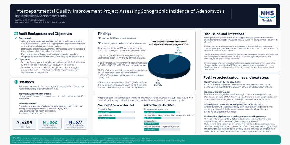
677 relevant TVUS reports were reviewed
577 scans suggested a diagnosis of adenomyosis
Two-thirds (66.2%, n = 382) of positive reports cited 2 to 3 sonographic features, detailed below
Only 16.8% (n = 97) relied on a single descriptor, whereas 83.0% (n = 479) cited 2 or more features.
Majority of patients were referred from primary care (80.2%, n=543/677 vs 19.8% from secondary care).
79.0% of all reviewed US requests did not include a specific clinical question of adenomyosis (n=538/677), suggesting high operator vigilance during TVUS.
MRI was performed in 20 out of 677 (3.0%) patients. This confirmed adenomyosis in 12 out of 16 patients and excluded adenomyosis in 3 out of 4 patients.
Morphological Uterus Sonographic Assessment (MUSA)2 consensus was first published in 2015 with the aim to refine diagnostic criteria and standardise ultrasound reporting for adenomyosis. See statistics attached.
Discussion and limitations:
Although not directly comparable, results suggest relative alignment with estimated disease burden provided by NICE guidelines (2018, 2021)3 in a symptomatic population at 20–35%.
Clinical notes were not reviewed due to the scope of project, high case volume and resource limitations. There was also no specific review of the number or type of presenting symptom(s) on the imaging requests.
Unclear how many patients subsequently received further gynaecological review or a final clinical diagnosis of adenomyosis. However, we noted a high TVUS sensitivity and specificity on patients followed up with MRI.
Common usage of legacy terms like “heterogeneous myometrium” reflect myometrial changes now better defined by MUSA terminology (eg. hyperechoic islands).
Local reporting practice is expected to gradually evolve towards these standardised descriptors.
Positive project outcomes and next steps:
1. High TVUS sensitivity and specificity:
Provided robust diagnostic certainty, eliminating the need for routine confirmatory pelvic MRI in the absence of additional clinical indications.
2. High reporting standards:
Feedback to sonographers and radiologists at our meeting reinforced standardised usage of MUSA terminology, therefore minimising variations in care and improving diagnostic pathways in line with national priorities.
3. Second phase retrospective analysis of this patient cohort:
Ongoing work with the gynaecology team to ascertain the proportion of patients reviewed clinically following imaging and if a final clinical or pathological diagnosis was made.
4. Optimisation of primary-secondary care diagnostic pathways:
Clinically mild or incidentally detected adenomyosis may be managed conservatively without requiring secondary care referral.
Our ongoing data collection on clinical symptoms and management would help risk stratify patients who would benefit from early gynaecology referral.
These insights will be fed back to primary care to enhance GP engagement and explore the use of a standardised patient symptom questionnaire.
References:
1. Taylor MA, Croudace TJ, Muir FE, McBride M. Women's experiences of living with adenomyosis and perceptions of the diagnostic journey: a scoping review. BMJ Open. 2025;15(1):e087122. doi:10.1136/bmjopen-2024-087122
2. Harmsen MJ, Van den Bosch T, de Leeuw RA, et al. Consensus on revised definitions of Morphological Uterus Sonographic Assessment (MUSA) features of adenomyosis: results of modified Delphi procedure. Ultrasound Obstet Gynecol. 2022;60(1):118–131. doi:10.1002/uog.24786
3. National Institute for Health and Care Excellence (NICE), 2018. Heavy menstrual bleeding: assessment and management (NICE Guideline NG88). Last updated May 2021. Available at: nice.org.uk