383 posters, 2 videos, 1 audios, 8 topics, 28 sessions, 328 authors, 225 institutions
ePostersLive by SciGen Technologies S.A. All rights reserved.
50th Annual Regional Anesthesiology and Acute Pain Medicine Meeting
May 1 - 3, 2025 | Orlando, Florida

6435
CT EVIDENCE OF LUMBAR SPINAL CANAL INCREASE POST DECOMPRESSION; A CASE REPORT
Poster Presenter
Authors
Affiliations
Part of Topic
Scientific Abstracts > Chronic Pain
CT Evidence of Lumbar Spinal Canal Increase Post Decompression; A Case Report
Victoria Portillo, Hyun Hong, Hannah Hong
Washington Center for Pain Management
Discussion
This case highlights several key clinical insights:
-
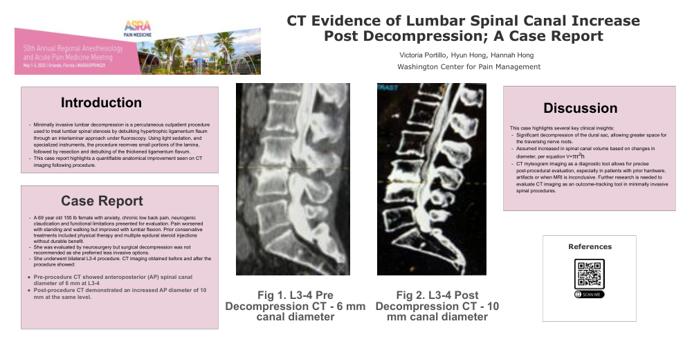
Significant decompression of the dural sac, allowing greater space for the traversing nerve roots.
-
Assumed increased in spinal canal volume based on changes in diameter, per equation V=πr2h
-
CT myleogram imaging as a diagnostic tool allows for precise post-procedural evaluation, especially in patients with prior hardware, artifacts or when MRI is inconclusive. Further research is needed to evaluate CT imaging as an outcome-tracking tool in minimally invasive spinal procedures.
Introduction
-
Minimally invasive lumbar decompression is a percutaneous outpatient procedure used to treat lumbar spinal stenosis by debulking hypertrophic ligamentum flaum through an interlaminar approach under fluoroscopy. Using light sedation, and specialized instruments, the procedure reomves small portions of the lamina, followed by resection and debulking of the thickened ligamentum flavum.
-
This case report highlights a quantifiable anatomical improvement seen on CT imaging following procedure.
Case Report
-
A 69 year old 155 lb female with anxiety, chronic low back pain, neurogenic claudication and functional limitations presented for evaluation. Pain worsened with standing and walking but improved with lumbar flexion. Prior conservative treatments included physical therapy and multiple epidural steroid injections without durable benefit.
-
She was evaluated by neurosurgery but surgical decompression was not recommended as she preferred less invasive options.
-
She underwent bilateral L3-4 procedure. CT imaging obtained before and after the procedure showed:
-
Pre-procedure CT showed anteroposterior (AP) spinal canal diameter of 6 mm at L3-4
-
Post-procedure CT demonstrated an increased AP diameter of 10 mm at the same level.
References
Fig 1. L3-4 Pre
Decompression CT - 6 mm canal diameter
Fig 2. L3-4 Post
Decompression CT - 10 mm canal diameter