425 posters, 1 videos, 9 audios, 8 topics, 53 sessions, 340 authors, 286 institutions
ePostersLive by SciGen Technologies S.A. All rights reserved.
22nd Annual Pain Medicine Meeting 2023
November 10 - 11, 2023 | New Orleans Marriott | New Orleans, Louisiana, USA

4506
AXIAL LOW BACK PAIN COMPLICATED BY MAY-THURNER SYNDROME WITH SPINAL CANAL STENOSIS: A CASE REPORT
Poster Presenter
Authors
Affiliations
Part of Topic
Chronic Pain
Axial low back pain complicated by May-Thurner Syndrome with spinal canal stenosis
Introduction
May-Thurner syndrome (MTS) is a relatively rare anatomical condition in which iliocaval venous territory is compressed by the arterial system against bony structures, most often the left common iliac vein by the right common iliac artery.
Patient
A 74-year-old male with a history of MTS status post left common iliac vein stent and aortofemoral artery bypass was seen in an outpatient pain management clinic with axial low back and referred hip pain
Case Description/Results
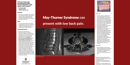
MRI thoracic and lumbar spine demonstrated extensive multilevel degenerative changes and facet arthrosis with severe central canal stenosis at T10-11 and throughout the lumbar spine from degenerative disc disease, ligamentum flavum thickening, and epidural fat prominence with noted epidural venous plexus engorgement throughout the thoracic and lower lumbar spine. Given the extensive degenerative findings, it was determined that surgical intervention would require a large thoracolumbar fusion and the patient opted for conservative management. Physical exam showed limited range of motion of the spine and provoked pain with facet-loading in the lumbar facet joints without radiculopathy, myelopathy, or neurogenic claudication. He underwent bilateral L3-5 medial branch blocks and reported 100% relief of pain. Subsequently, he underwent successful right L3-5 medial branch radiofrequency (RFA) but was unable to tolerate left-sided RFA due to discomfort with needle positioning.
Discussion
This case shows the importance of advanced imaging, careful physical exam, and appropriate vascular workup in patients with MTS. Patients with MTS often present with extremity pain and swelling, venous claudication with thigh and leg pain, and persistent groin or low back pain; these symptoms may mimic common causes of mechanical low back and radicular pain. Engorgement of epidural vessels may lead to compression of nerves, contributing to lumbosacral radiculopathy or even cauda equina syndrome. In this case, the patient presented with symptoms that appear common in patients with chronic low back pain after appropriate treatment of MTS. It is possible that despite previous vascular treatment of MTS, his vascular symptoms contributed to increased pain and worse tolerance of intervention on the left. Given the success of the diagnostic and confirmatory medial branch blocks, repeat RFA may be attempted with appropriate sedation for improved procedural pain tolerance.
Conclusion
Special attention should be given to advanced imaging that may lead to epidural venous plexus engorgement, which may contribute to significant spinal canal stenosis, symptoms of neurogenic claudication, and low back pain.