425 posters, 1 videos, 9 audios, 8 topics, 53 sessions, 340 authors, 286 institutions
ePostersLive by SciGen Technologies S.A. All rights reserved.
22nd Annual Pain Medicine Meeting 2023
November 10 - 11, 2023 | New Orleans Marriott | New Orleans, Louisiana, USA

4505
DYNAMIC INSTABILITY IS UNDERESTIMATED ON STANDING FLEXION-EXTENSION FILMS WHEN COMPARED TO PRONE CT IMAGING
Poster Presenter
Authors
Affiliations
Part of Topic
Chronic Pain
Introduction
Dynamic lumbar instability evaluated on flexion-extension radiographs has been a longstanding convention in the decision-making for identifying the level of axial and radicular pain and its consequent management. However, flexion-extension radiographs have been shown to underestimate the degree of lumbar spondylolisthesis. Significant variability remains in current guidelines regarding the most appropriate imaging modalities to adequately evaluate instability. We hypothesize that prone positioning will provide greater sensitivity for dynamic instability compared to standing or supine positions. While prior studies have instability with either supine CT or MRI, this is the first study to our knowledge measuring single level lumbar instability with prone positioning obtained during interventional spinal injections.
Methods
▪ Single center retrospective cohort study of single-level (L4-5) anterolisthesis between 2014 – 2022 with standing radiographs (CR), prone CT images (CT), or supine MRI images (MRI)
▪ Exclusion Criteria: Prior lumbar hardware, missing imaging modalities time between any modalities > 13 months, no evidence of anterolisthesis
▪ Anterior translation was measured in millimeters across imaging modalities between two raters, and the translation compared across different modalities.
▪ We calculated a ratio which was the amount of translation divided by the width of the vertebral body for each image modality (CR, CT, or MRI) and multiplied this by the width of the vertebral body measured on MRI.
Results
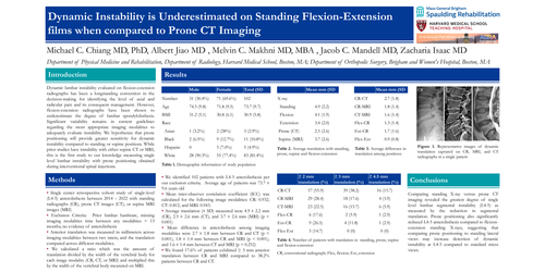
▪ We identified 102 patients with L4-5 anterolisthesis per our exclusion criteria. Average age of patients was 73.7 ± 9.6 years old
▪ Mean inter-observer correlation coefficient (ICC) was calculated for the following image modalities: CR: 0.932; CT: 0.812; and MRI: 0.943.
▪ Average translation (± SD) measured were 4.9 ± 2.2 mm (CR), 2.5 ± 2.6 mm (CT), and 3.7 ± 2.6 mm (MRI) (p < 0.001)
▪ Mean difference in anterolisthesis among imaging modalities were 2.7 ± 1.8 mm between CR and CT (p < 0.001), 1.8 ± 1.4 mm between CR and MRI (p < 0.001), and 1.6 ± 1.4 mm between CT and MRI (p = 0.252)
▪ We found 17.6% of patients exhibited ≥ 3 mm anterior translation between CR and MRI compared to 38.2% patients between CR and CT.
Conclusion
Comparing standing X-ray versus prone CT imaging revealed the greatest degree of single level lumbar segmental instability (L4-5) as measured by the reduction in segmental translation. Prone positioning also significantly reduced L4-5 anterolisthesis compared to flexion-extension standing X-rays, suggesting that comparing prone positioning to standing lateral views may increase detection of dynamic instability at L4-5 compared to standard stress views.