425 posters, 1 videos, 9 audios, 8 topics, 53 sessions, 340 authors, 286 institutions
ePostersLive by SciGen Technologies S.A. All rights reserved.
22nd Annual Pain Medicine Meeting 2023
November 10 - 11, 2023 | New Orleans Marriott | New Orleans, Louisiana, USA

4718
WORSENING RETROPULSION OF BONY FRAGMENTS FOLLOWING IMAGE GUIDED RADIOFREQUENCY ABLATION AND VERTEBROPLASTY
Poster Presenter
Authors
Affiliations
Part of Topic
Chronic Pain
Worsening Retropulsion of Bony Fragments Following Image Guided Radiofrequency Ablation and Vertebroplasty
Percutaneous vertebroplasty is commonly indicated for the treatment of medically refractory vertebral body compression fractures. • Relative contraindications include disruption of the posterior vertebral body wall or tumor extension into the spinal canal. • Radiofrequency ablation (RFA) can be incorporated to improve pain outcomes. • Complications may arise even after reassuring post-operative imaging.
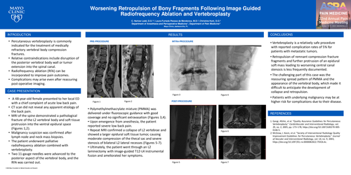
A 48-year-old female presented to her local ED with a chief complaint of acute low back pain. • CT scan did not reveal any apparent etiology of the back pain. • MRI of the spine demonstrated a pathological fracture of the L2 vertebral body and soft tissue protrusion into the ventral epidural space (Figures 1,2). • Malignancy suspicion was confirmed after lymph node and neck mass biopsies. • The patient underwent palliative radiofrequency ablation combined with vertebroplasty. • Two 11-gauge needles were advanced to the posterior aspect of the vertebral body, and the RFA was carried out. CASE PRESENTATION • Polymethylmethacrylate mixture (PMMA) was delivered under fluoroscopic guidance with good coverage and no significant extravasation (Figures 3,4). • Upon emergence from anesthesia, the patient reported severe low back pain. • Repeat MRI confirmed a collapse of L2 vertebrae and showed a larger epidural soft tissue tumor, causing moderate compression of the thecal sac and severe stenosis of bilateral L2 lateral recesses (Figures 5-7). • Ultimately, the patient went through an L2 laminectomy with image-guided T12-L4 instrumental fusion and ameliorated her symptoms.