425 posters, 1 videos, 9 audios, 8 topics, 53 sessions, 340 authors, 286 institutions
ePostersLive by SciGen Technologies S.A. All rights reserved.
22nd Annual Pain Medicine Meeting 2023
November 10 - 11, 2023 | New Orleans Marriott | New Orleans, Louisiana, USA

4819
IDIOPATHIC SAPHENOUS NERVE ENTRAPMENT: A CASE REPORT
Poster Presenter
Authors
Affiliations
Part of Topic
Chronic Pain
Idiopathic Saphenous Nerve Entrapment: A Case ReportWilliam Tang, M.D., Nikolle Lambrinos, M.D., Ly Vu, M.D., Danielle Horn, M.D.Department of Anesthesiology, Perioperative Medicine and Pain ManagementUniversity of Miami / Jackson Memorial Health System
INTRODUCTION:
The saphenous nerve is a branch of the femoral nerve that provides sensory innervation to the skin of the anteromedial leg. Saphenous nerve entrapment may result in saphenous neuropathy or neuralgia, a condition in which the saphenous nerve becomes compressed or irritated, leading to pain, numbness, or paresthesia along its distribution.[1] Saphenous nerve injury is relatively uncommon, amounting for less than 1% of adult patients with lower extremity pain. [2]
Potential causes of saphenous nerve entrapment include trauma, compression, anatomical anomalies, inflammatory conditions, and most commonly post-surgical complications (mainly surgeries in the knee or upper leg area). Neuralgia and neuroma formation may occur after iatrogenic saphenous nerve injury, such as after orthopedic surgeries or saphenectomy for vascular procedures.[3]
Symptoms of saphenous nerve entrapment include pain with or without associated numbness and tingling. The most common symptom is pain along the inner aspect of the knee and the lower leg. The distribution of pain in patients with saphenous nerve entrapment has been reported at the knee (90%), thigh (7%) and calf (3%).[4] The pain can range from mild to severe and may be described as burning, shooting, or throbbing.
The diagnosis of saphenous nerve entrapment typically involves a thorough medical history and physical examination. Imaging studies, such as MRI or ultrasound, may be limited. Nerve conduction studies may be utilized to assess the function of the nerve and to identify the site of entrapment. In addition, local anesthetics may be utilized as a diagnostic and/or therapeutic tool.[1]
This case report will review a patient who presented with idiopathic saphenous nerve entrapment diagnosed after a saphenous nerve (adductor canal) block.
CONSENT:
Patient informed consent was obtained for submission of this case report.
CASE DESCRIPTION:
A 59 year-old female with a medical history of hypertension and anxiety/depression presented with pain located along the medial aspect of her right thigh. The pain started approximately one year before her initial pain clinic consultation. Pain occurred mainly with abduction of her right leg and was described as an intermittent electrical sensation in this area. She experienced difficulty with crossing her right leg over her left knee, stating that she had to use her arms to assist her. She denied pain in her groin and any triggering factors or inciting events. Trials of gabapentinoids and non-steroidal anti-inflammatory drugs were unsuccessful. She was evaluated by orthopedic surgery and sports medicine specialists, and no surgery was recommended. Her physical exam was remarkable for tenderness along the medial aspect of the distal right thigh and pain initiated with abduction of the right lower extremity. MRI of the right femur did not show any soft tissue masses encroaching around the area of patient’s pain.
The top differential diagnosis considered was saphenous nerve entrapment, thus a right saphenous nerve block was performed under ultrasound guidance with the patient’s consent.
METHODS:
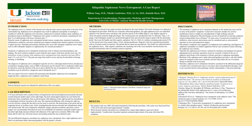
The patient was placed in the supine position and draped in the usual fashion. Full sterile technique was adhered to throughout the procedure. With the use of dynamic ultrasound guidance, the right saphenous nerve was identified adjacent to the femoral artery and deep to the sartorius muscle in the middle thigh at a site slightly superior to where the patient noted her thigh pain began. The overlying skin was anesthetized with 1% lidocaine. Then, a 21-gauge, 4-inch Stimuplex needle was advanced through the skin and toward the target in-plane with the ultrasound probe. Once near the bundle and after negative aspiration, 5ml of a 10ml of a solution containing ropivacaine 0.5% x4.5ml, preservative-free normal saline x 4.5ml, and dexamethasone 10mg/ml x1ml were injected incrementally with no resistance noted on injection. The patient did not report any paresthesias. The needle was redirected above the saphenous nerve. After negative aspiration, the remaining 5ml of the local anesthetic-steroid mixture was injected incrementally with no resistance noted on injection.
RESULTS:
•
The patient’s pain was 100% alleviated immediately following the procedure, with a pain score decreased from 7/10 to 0/10; and relief lasted approximately 5 months.
•
When the pain returned, the patient was scheduled for a repeat right adductor canal nerve block.
•
Approximately 7 months later, the patient reported continued improvement of her pain with about 90% resolution.
DISCUSSION:
•
The treatment of saphenous nerve entrapment depends on the underlying cause and the severity of the patient’s symptoms. Conservative measures include rest, activity modification (such as weight loss and adjustment of tight clothing), and anti-inflammatory medications. Physical therapy can also be beneficial in relieving symptoms and preventing further nerve irritation.[5] In some cases, if conservative treatments are ineffective, more invasive interventions may be considered such as corticosteroid injections or nerve decompression surgery.
•
This patient’s symptoms were suggestive of saphenous nerve pathology, and diagnosis of saphenous neuropathy was further supported with her near resolution of pain following the saphenous nerve blocks.
•
Typically, the saphenous nerve block is utilized for anesthesia and analgesia for patients undergoing lower extremity operations; however, research is limited on the use of adductor canal blocks in the treatment of chronic pain due to saphenous neuropathy.
•
Blockade of the saphenous nerve under ultrasound guidance provides a safe and reliable option for analgesia of the lower extremity and may help reduce the use of systemic therapy in patients with severe pain.
•
Therefore, saphenous nerve entrapment should be considered in the differential diagnosis of medial lower extremity pain since it can mimic other musculoskeletal disorders, such as pes anserinus bursitis, knee pathology, lumbar radiculopathy, or hip joint pathology.
REFERENCES:
1.
Morganti, Christina M et al. “Saphenous neuritis: a poorly understood cause of medial knee pain.” The Journal of the American Academy of Orthopaedic Surgeons vol. 10,2 (2002): 130-7. doi:10.5435/00124635-200203000-00008
2.
Mumenthaler, Marco, Hans Schliack, and Heinz Goerke. Peripheral nerve lesions: diagnosis and therapy. Thieme Medical Publishers, 1991.
3.
Kachar, Sergey M., Kristopher M. Williams, and Henry A. Finn. "Neuroma of the infrapatellar branch of the saphenous nerve: a cause of reversible knee stiffness after total knee arthroplasty." The Journal of arthroplasty 23.6 (2008): 927-930.
4.
Romanoff, M E et al. “Saphenous nerve entrapment at the adductor canal.” The American journal of sports medicine vol. 17,4 (1989): 478-81. doi:10.1177/036354658901700405
5.
Settergren, Roy. “Conservative management of a saphenous nerve entrapment in a female ultra-marathon runner.” Journal of bodywork and movement therapies vol. 17,3 (2013): 297-301. doi:10.1016/j.jbmt.2012.10.005