203 posters, 14 topics, 5 sessions, 817 authors, 256 institutions
ePostersLive by SciGen Technologies S.A. All rights reserved.
The AAPS 102nd Annual Meeting
May 18-21, 2024 | The Westin Copley Place, Boston, MA

D2
Mixed Metal Oxide Nanoparticles For Seroma Treatment: A Comparative Long-term Study In Rats
Poster Presenter
Part of Topic
Basic Science Research
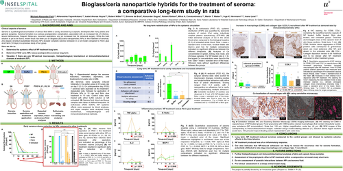
Bioglass/ceria nanoparticle hybrids for the treatment of seroma: a comparative long-term study in rats
1 Department of Plastic and Hand Surgery, Inselspital, University Hospital Bern, Bern, Switzerland; 2 Department for BioMedicalResearch, University of Bern, Bern, Switzerland; 3Department of Materials Meet Life, Swiss Federal Laboratories for Materials Science and Technology (Empa), St. Gallen, Switzerland; 4 Department of Mechanical and Process Engineering, ETH Zurich, Zurich, Switzerland; 5 Ingenuity Lab, University Hospital Balgristand University of Zurich, Zurich, Switzerland; 6COMPATH, Institute of Animal Pathology, University of Bern, Bern, Switzerland
1. BACKGROUND AND AIMS
Clinical aspects of seroma:
Seroma is a pathological accumulation of serous fluid within a cavity, enclosed by a capsule, developed after many plastic and general surgeries. Seroma formation is a serious postoperative complication, associated with an increased risk of infection, wound dehiscences, frequent follow-ups, recurrent aspirations, and an increased risk for revisional surgery. While the best solution is yet to be found, recent focus has been put on Bioglass (BG)/ceria nanoparticles (NPs) in the treatment of seromas. Zinc-doped, strontium-substituted BG/ceria NPs showed complete seroma remission in a rat model compared to Fibrin glue short term (14 days) in a previous study of our group.
Here we aim to:
1.Determine the systemic effect of NP treatment long term.
2. METHODS
Fig. 1. Experimental design for seroma induction, formation, aspiration, and treatments in Lewis rats (n=20). (A) Seromas were surgically induced bilaterally in the axillary area of 20 Lewis rats on day 0 (D 0). On postoperative day (POD) 7 seromas were aspirated on the treatment-designated side, followed by application of BG/ceria NPs in 10 rats and fibrin glue treatment in 10 rats. Control sides were treated with either buffer solution or left untreated. (B) Seroma fluid, blood and tissue samples were taken at defined timepoints. At euthanasia (POD 42/EP), NP systemic effects were assessed by blood analyses, and organ samples were analysed using biochemical, histopathological and immunohistochemical methods.
3. RESULTS
Early seroma volume reduction by NPs compared to other treatments
Fig. 2. (A) After seroma fluid aspiration on POD 7, the treatment sides were injected with either NPs or fibrin glue. At PODs 14, 21, 28, 35, and 42, seroma-fluid volumes were assessed after aspiration, and indicated as percentage of the recorded volume [VOLpct] (B) NP treatment resulted in significant early fluid reduction at POD 14. Data = mean ± standard error of the mean. ****p < 0.0001, NPs and buffer solution vs untreated at POD 14. *p = 0.0176 and NPs vs fibrin glue, respectively *p = 0.0219, buffer solution vs fibrin glue at POD 14. Two-way ANOVA with Tukey post hoc for multiple comparisons.
No long-term redistribution of NPs to the systemic circulation
Fig. 3 At euthanasia (POD 42), systemic distribution of NPs was quantified by elemental analysis of cerium (Ce) using inductively coupled plasma spectroscopy (ICP-MS). (A) Initial elemental analysis of Ce in the whole blood when compared to the biological baseline (D 0) of untreated whole blood. Data = mean ± standard error of the mean. Kruskal–Wallis with Dunn`s post hoc for multiple comparisons indicated no significant differences between the different timepoints. (B) Analyses of Ce concentration in spleen, kidney, liver, brain, and LN tissue samples. For NP-treated rats (n = 9), the largest Ce concentrations were found in the liver. Data = mean ± standard error of the mean. Wilcoxon tests without significant differences between the groups.
Long term, NP treatment led to cavity reductions and increased adhesions
Fig. 4 (A) At endpoint (POD 42), the surgical seroma sites were scored for macroscopic changes, including cavities and single/multiple adhesions with either focal or planar attachments. Scoring ranged from 0–3 points, with 0 representing no adhesions, but a cavity, and 3 representing multiple adhesions. (B) Complete resolution of cavities was observed in 5 out of 9 NP-treated rats. Data = mean ± standard error of the mean. Kruskal–Wallis with Dunn`s post hoc for multiple comparisons. ***p = 0.0004, NP vs untreated, and *p = 0.0358, NP vs fibrin glue.
Inflammatory markers: NP treatment versus fibrin glue treatment
Fig. 5 (A-D) Quantitative assessments of plasma analytes using a commercial kit (Luminex, BioPlex). When pg/mL values were not detectable (<3.17 for TNF-alpha, <9.45 for IL-1beta, <1.68 for IL-2, and <14.7 for MCP-1) they were assigned a bin value of 0. Data = mean ± standard error of the mean. Significant differences highlighted in red-framed close ups at the respective timepoints. *p = 0.028, TNF- alpha at POD 42, *p = 0.0294, IL1-beta at POD 14, *p = 0.014, IL-2 at POD 14, **p = 0.0048, MCP-1 at POD 28, NPs vs fibrin glue. Mann- Whitney tests for single comparisons. Two-way ANOVA with Bonferroni post hoc for multiple comparisons indicated no significant differences between the different treatments.
Increase in macrophage (CD68) and collagen type I (COL1) recruitment after NP treatment as demonstrated by immunohistochemistry (IHC)
Fig. 6 IHC staining for CD68, COL1, containing the superficial seroma capsule of NP treated, buffer treated, fibrin glue treated, and untreated groups. Overview and enlarged areas at the capsular level of skin tissue sections. Scale bars, 1 mm. (A) CD68. Nodular dense aggregates of CD68 positive cells correspond to granulomas which are most extensive after NP, and absent in the untreated side. (B) COL1. Collagen deposition tended to be multifocal and to be denser after NP treatment compared to other treatment groups.
Fig. 7 Quantitative assessments of IHC staining for CD68, CD31 and COL1 in capsule tissue. (A) Increased granuloma formation (grouped macrophages) after NP treatment. Area of CD68 positive stained granuloma was measured and indicated as percentage of the assessed capsule tissue [AApct]. Data = mean ± standard error of the mean. (B) Increases in more intense COL1 deposition after NP treatment. IHC staining for COL1 measuring the ratio between more vs less intensely stained (i.e., densely arranged) collagen [more intensity/less intensity]. Data = mean ± standard error of the mean. Mann-Whitney tests. **p = 0.0221, NP vs untreated, Mann-Whitney tests for single comparisons.
Co-localization of macrophages with NPs using correlative microscopy
Fig. 8 Correlation between IHC and Scanning Electron Microscopy (SEM) imaging techniques. (A) IHC staining for CD68 in capsule tissue of NP treated groups. Focal granuloma with CD68 positive stained macrophages that contain intracellular purple-grey substance (NP accumulation). Scale bar, 100 μm. Higher magnification, scale bar, 40 μm. (B) SEM images of the corresponding to immunohistochemical images, collected using a backscattering detector (A.). Electron dense region sections (scale bars, 100 μm) and maps indicating cerium represented in pink color (Ce).
4. CONCLUSIONS
5. FUTURE DIRECTIONS
6. ACKNOWLEDGEMENTS
The project is partially funded by an Innosuisse grant. (Project no.: 54684.1 IP-LS).