203 posters, 14 topics, 5 sessions, 817 authors, 256 institutions
ePostersLive by SciGen Technologies S.A. All rights reserved.
The AAPS 102nd Annual Meeting
May 18-21, 2024 | The Westin Copley Place, Boston, MA

P35
Quantifying Type H Vasculature In Irradiated Distraction Osteogenesis As A Marker For Enhanced Bone Healing
Poster Presenter
Part of Topic
Hand / Craniofacial
Quantifying Type H Vasculature In Irradiated Distraction Osteogenesis As A Marker For Enhanced Bone Healing
Melissa Daniel MD MPH1, Nathan Sheppard B Arch2, Noah Nelson BS MPH2, Alex Donneys MD2, Steven Buchman MD2
1University of Oklahoma-Tulsa, Tulsa, OK
2Craniofacial Research Laboratory, University of Michigan, Ann Arbor, MI
Study Goals
Methods
Results
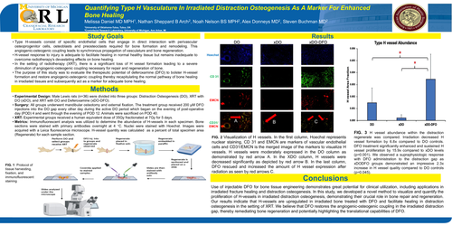
FIG. 2 Visualization of H vessels. In the first column, Hoechst represents nuclear staining, CD 31 and EMCN are markers of vascular endothelial cells and CD31/EMCN is the merged image of the markers to visualize H vessels. H vessels were moderately expressed in the DO column as demonstrated by red arrow A. In the XDO column, H vessels were decreased significantly as depicted by red arrow B. In the last column, DFO rescued and increased the amount of H vessel expression after radiation as seen by red arrows C.
FIG. 3 H vessel abundance within the distraction regenerate was compared. Irradiation decreased H vessel formation by 6.8x compared to DO controls. DFO treatment significantly enhanced and sustained H vessel proliferation by 15.9x compared to xDO levels (p=0.001). We observed a supraphysiologic response with DFO administration to the distraction gap as xDODFO groups demonstrated an impressive 2.3x increase in H vessel quality compared to DO controls (p=0.045).
Conclusions
Use of injectable DFO for bone tissue engineering demonstrates great potential for clinical utilization, including applications in irradiated fracture healing and distraction osteogenesis. In this study, we developed a novel method to visualize and quantify the proliferation of H-vessels in irradiated distraction osteogenesis, demonstrating their crucial role in bone repair and regeneration. Ourresults indicate that H-vessels are upregulated in irradiated bone treated with DFO and facilitate healing in distraction osteogenesis in the setting of XRT. We believe that DFO restores the angiogenic-osteogenic coupling in the irradiated distraction gap, thereby remediating bone regeneration and potentially highlighting the translationalcapabilities of DFO.