203 posters, 14 topics, 5 sessions, 817 authors, 256 institutions
ePostersLive by SciGen Technologies S.A. All rights reserved.
The AAPS 102nd Annual Meeting
May 18-21, 2024 | The Westin Copley Place, Boston, MA

P31
Immune Repertoire In Lymphedema: Characterizing The Dendritic Cell Subsets In A Lymphedema Mouse Model
Part of Topic
Hand / Craniofacial
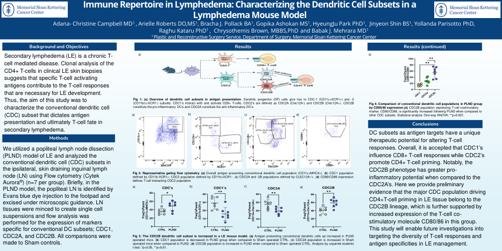
Immune Repertoire in Lymphedema: Characterizing the Dendritic Cell Subsets in a Lymphedema Mouse Model
Secondary lymphedema (LE) is a chronic T-cell mediated disease. Clonal analysis of the CD4+ T-cells in clinical LE skin biopsies suggests that specific T-cell activating antigens contribute to the T-cell responses that are necessary for LE development. Thus, the aim of this study was to characterize the conventional dendritic cell (CDC) subset that dictates antigen presentation and ultimately T-cell fate in secondary lymphedema.
We utilized a popliteal lymph node dissection (PLND) model of LE and analyzed the conventional dendritic cell (CDC) subsets in the ipsilateral, skin draining inguinal lymph node (LN) using Flow cytometry (Cytek Aurora®) (n=7 per group). Briefly, in the PLND model, the popliteal LN is identified by Evans blue dye injection to the footpad and excised under microscopic guidance. LN tissues were minced to create single cell suspensions and flow analysis was performed for the expression of markers specific for conventional DC subsets; CDC1, CDC2A, and CDC2B. All comparisons were made to Sham controls.
Fig 1: (a) Overview of dendritic cell subsets in antigen presentation. Dendritic progenitor (DP) cells give rise to CDC-1 (CD11c+XCR1+) and -2 (CD11b/c+XCR1-) subsets. CDC1’s interact with and activate CD8+ T-cells. CDC2’s are defined as CDC2A (Clec12A-) and CDC2B (Clec12A+). CDC2B constitute the pro-inflammatory DC’s and CDC2A constitute the anti-inflammatory.
Fig 2: Representative gating flow cytometry. (a) Overall antigen presenting conventional dendritic cell population (CD11c+MHCII+). (b) CDC1 population defined by CD11b-XCR1+; CDC2 population defined by CD11b+XCR1-. (c) CDC2A and -2B populations defined by CLEC12A-/+. (d) CD80/CD86 expression defines T-cell interacting CDC2 population
Fig 3: The CDC2B dendritic cell subset is increased in a LE mouse model. (a) Antigen presenting conventional dendritic cells ae increased in PLND operated mice. (b) CDC1 population is decreased in PLND group when compared to CTRL. (c) CDC2A population is increased in Sham operated mice when compared to PLND. (d) CDC2B population is increased in PLND when compared to Sham operated CTRL. Analysis by unpaired students t-test; *p<0.05, **p<0.001
DC subsets as antigen targets have a unique therapeutic potential for altering T-cell responses. Overall, it is accepted that CDC1’s influence CD8+ T-cell responses while CDC2’s promote CD4+ T-cell priming. Notably, the CDC2B phenotype has greater pro-inflammatory potential when compared to the CDC2A’s. Here we provide preliminary evidence that the major CDC population driving CD4+T-cell priming in LE tissue belong to the CDC2B lineage, which is further supported by increased expression of the T-cell co-stimulatory molecule CD80/86 in this group. This study will enable future investigations into targeting the diversity of T-cell responses and antigen specificities in LE management.