203 posters, 14 topics, 5 sessions, 817 authors, 256 institutions
ePostersLive by SciGen Technologies S.A. All rights reserved.
The AAPS 102nd Annual Meeting
May 18-21, 2024 | The Westin Copley Place, Boston, MA

D157
Intraoperative Indocyanine Green Angiography For Detecting Quantitative Flap Perfusion Changes In Deep Inferior Epigastric Perforator Flap Breast Reconstruction With Second Venous Drainage
Part of Topic
Breast Reconstruction
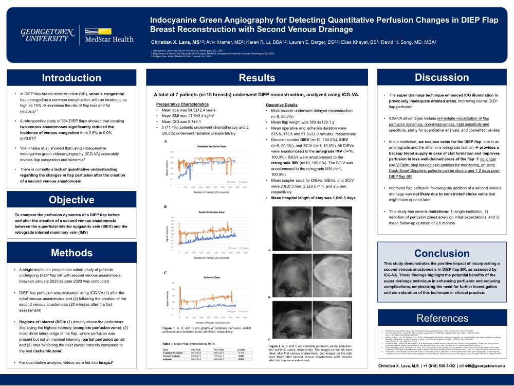
Indocyanine Green Angiography for Detecting Quantitative Perfusion Changes in DIEP Flap Breast Reconstruction with Second Venous Drainage
Background: Venous congestion poses a risk of necrosis and complete flap loss in deep inferior epigastric perforator (DIEP) flap breast reconstructions. To address this issue, some plastic surgeons have started incorporating two venous anastomoses. However, there is a need for a quantitative understanding of the changes in flap perfusion following the second anastomosis. This study aims to compare perfusion dynamics using indocyanine green videoangiography (ICG-VA) before and after the creation of a second venous anastomosis between the superficial inferior epigastric vein (SIEV) and retrograde internal mammary vein (IMV).
Methods: ICG-VA performed during DIEP flap reconstructions was analyzed prospectively. The angiogram was captured serially in a regular time interval. The stacked images were then fed into an image analysis program, ImageJ. The selected areas of interest were as follows: above the perforators with the highest intensity (complete perfusion zone), the most distal lateral edge of the flap (partial perfusion zone), and the next lowest intensity zone (ischemic zone). We compared the intensity of these zones before and after the second venous anastomosis, and venous drainage patency and functionality. Patient characteristics, preoperative characteristics, surgical technique employed, intraoperative details, and postoperative outcomes (e.g., complications) were collected.
Results: Seven patients underwent DIEP reconstruction, with a total of 10 breasts analyzed using ICG-VA. Mean age and BMI were 54.5±12.4 years and 27.6±3.4 kg/m 2 , respectively. Mean flap weight was 503.4±129.1 g. Mean operative and ischemia duration were 575.5±172.6 and 67.6±22.0 minutes, respectively. Donors included DIEV (n=10, 100.0%), SIEV (n=9, 90.0%), and superficial circumflex epigastric vein (SCIV) (n=1, 10.0%). All DIEVs were anastomosed to the antegrade IMV (n=10, 100.0%). SIEVs were anastomosed to the retrograde IMV (n=10, 100.0%). Mean peak intensity of the complete perfusion zone before and after second venous anastomosis were 160.7±42.1 and 188±42.1, respectively (p=0.163). Mean peak intensity of the partial perfusion zone before and after second venous anastomosis were 100.8±21.5 and 152±31.5, respectively (p<0.001). Mean peak intensity of the ischemic zone before and after second venous anastomosis were 90.4±37.4 and 143.4±45.3, respectively (p=0.012).
Conclusion: This study demonstrates the positive impact of incorporating a second venous anastomosis in DIEP flap breast reconstruction, as assessed by ICG-VA. These findings highlight the potential benefits of the super drainage technique in enhancing perfusion and reducing complications, emphasizing the need for further investigation and consideration of this technique in clinical practice.