212 posters, 10 topics, 5 sessions, 754 authors, 237 institutions
ePostersLive by SciGen Technologies S.A. All rights reserved.
The AAPS 101st Annual Meeting
29 April – 2 May, 2023 | Chicago, IL

PC46
MRI Neurography As An Adjunct For The Diagnosis Of Meralgia Paresthetica
Poster Presenter
Part of Topic
Outcomes
MRI Neurography as an Adjunct for the Diagnosis of Meralgia Paresthetica
Introduction
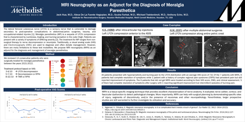
The lateral femoral cutaneous nerve (LFCN) is a sensory nerve that is vulnerable to damage secondary to post-operative complications in abdominal/pelvic surgeries, trauma, and occupational-related injuries [1]. Meralgia paresthetica (MP) is a sequela of LFCN compression that is characterized by numbness, tingling, and burning sensation in the outer thigh. Patients can present with a variety of symptoms of differing severity [3]. The treatment for MP ranges from non-surgical therapy to nerve decompression or neurolysis. Traditionally, a visual analog scale (VAS) and Electromyography (EMG) are used to diagnose and often dictate management. However, there are many limitations to these test modalities. We propose MRI neurography (MRN) as an adjunct for the diagnosis and management of meralgia paresthetica.
Methods
We reviewed 13 consecutive patients who were surgically treated for meralgia paresthetica between the years 2021-2022. Our treatment protocol offered patients with VAS scores of 3-4 with nerve release, 5-7 with either LFCN decompression or resection with regenerative peripheral nerve interface (RPNI), and 8-10 with RPNI. Since MRI neurography was only recently introduced into our practice, 7 out of the 13 patients had MRN pre-operatively.
Results:
All patients initially presented with hypersensitivity and burning pain to the LFCN distribution with an average VAS score of 7.8. Of the 7 patients with MRN, 6 patients had complete resolution of symptoms while 1 patient with a history of complex regional pain syndrome (CRPS) had persistent pain but still reported improvement of symptoms. The 6 patients that did not get MRN were treated according to their VAS score, EMG, and clinical assessment. 5 of them had complete resolution of symptoms after surgery with 1 reporting significant improvement but intermittent episodes of burning pain.
Conclusion
MRN is a tissue-specific imaging technique that provides excellent characterization of nerve anatomy. It evaluates nerve caliber, contour, and fascicular dysfunction to detect pathological changes. More importantly, MRN can help with surgical planning by demonstrating specific sites of injury, segments of discontinuity, and/or the presence of neuromas and other neuropathologies.