4,068 posters, 219 videos, 101 topics, 69 sessions, 14,365 authors, 6,128 institutions
ePostersLive by SciGen Technologies S.A. All rights reserved.
Alzheimer's Association International Conference 2026
July 12-15, 2026 | London, United Kingdom, and Online

10016
Longitudinal Multimodal Magnetic Resonance Imaging Analysis Of Lecanemab Treatment In Mild Cognitive Impairment: A Pilot Study
Part of Topic
Longitudinal Multimodal Magnetic Resonance Imaging Analysis of Lecanemab Treatment in Mild Cognitive Impairment: A Pilot Study
Toshiya Takahashi, Hidenao Fukuyama
Department of Neurology, Graduate School of Medicine, Kyoto University, Kyoto, Japan.
Human Brain Research Center, Kyoto University Graduate School of Medicine, Kyoto, Japan.
Background / Objective:
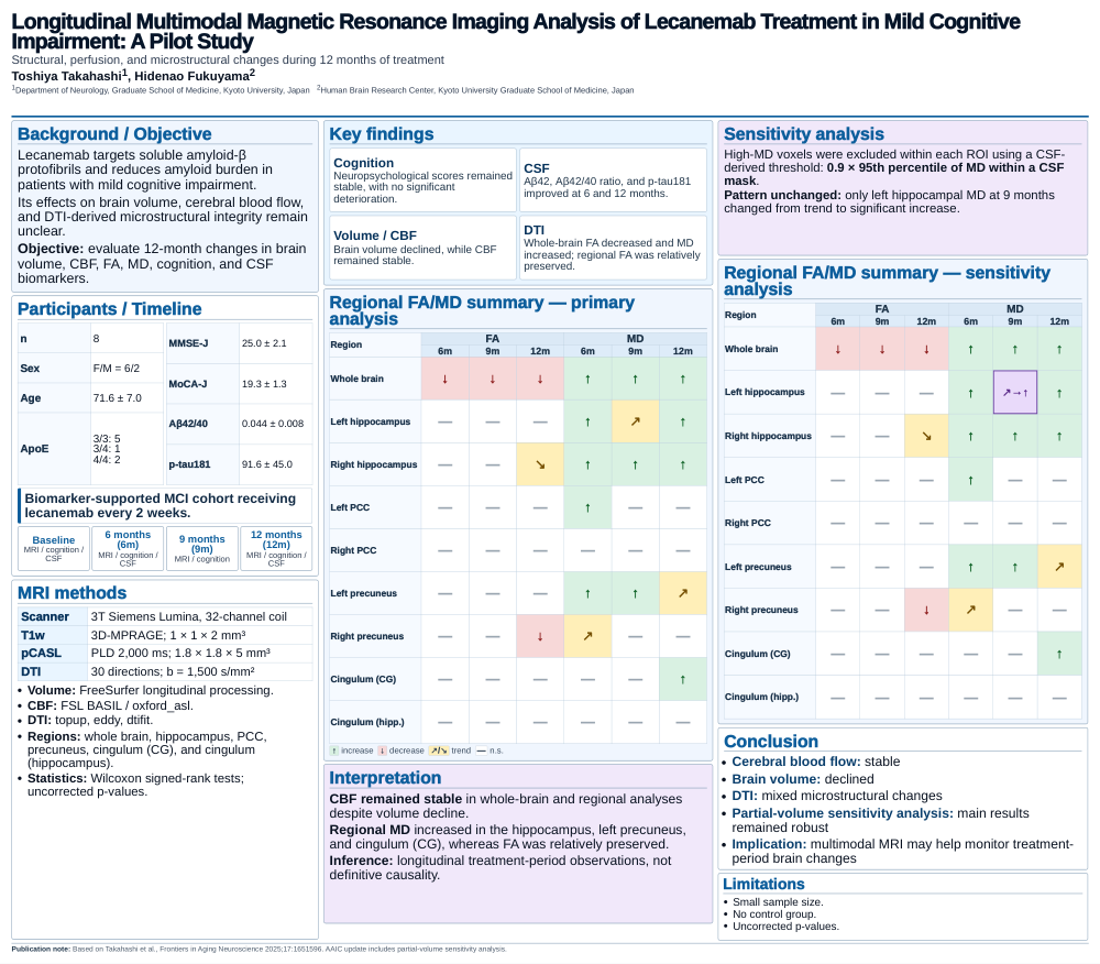
Lecanemab targets soluble amyloid-β protofibrils and reduces amyloid burden in patients with mild cognitive impairment. Its effects on brain volume, cerebral blood flow, and DTI-derived microstructural integrity remain unclear. This exploratory longitudinal study evaluated 12-month changes in brain volume, cerebral blood flow, fractional anisotropy, mean diffusivity, cognition, and cerebrospinal fluid biomarkers in patients with mild cognitive impairment receiving lecanemab treatment.
Participants / Timeline:
Eight patients with biomarker-supported mild cognitive impairment received lecanemab every 2 weeks. MRI, neuropsychological assessments, and cerebrospinal fluid biomarkers were assessed longitudinally. Imaging and cognitive assessments were performed at baseline, 6 months, 9 months, and 12 months. Cerebrospinal fluid biomarkers were assessed at baseline, 6 months, and 12 months.
MRI methods:
MRI was acquired using 3T Siemens Lumina with a 32-channel head coil. Structural MRI used 3D-MPRAGE. Cerebral blood flow was assessed using pseudo-continuous arterial spin labeling. Diffusion tensor imaging was used to calculate fractional anisotropy and mean diffusivity. Brain volume was analyzed using FreeSurfer longitudinal processing. Cerebral blood flow was analyzed using FSL BASIL / oxford_asl. DTI preprocessing included topup, eddy, and dtifit. Regions of interest included the whole brain, hippocampus, posterior cingulate cortex, precuneus, cingulum (cingulate gyrus), and cingulum (hippocampus). Statistical analyses used Wilcoxon signed-rank tests.
Key findings:
Neuropsychological scores remained stable, with no significant deterioration. Cerebrospinal fluid biomarkers improved, including Aβ42, Aβ42/40 ratio, and p-tau181 at 6 and 12 months. Brain volume declined in the whole brain and key Alzheimer’s disease-related regions. Cerebral blood flow remained stable in whole-brain and regional analyses. At the whole-brain level, fractional anisotropy decreased and mean diffusivity increased. In regional analyses, mean diffusivity increased in the hippocampus, left precuneus, and cingulum (cingulate gyrus), whereas fractional anisotropy was relatively preserved, suggesting region-dependent microstructural changes.
Partial-volume sensitivity analysis:
To address cerebrospinal fluid partial-volume effects, high-MD voxels were excluded within each ROI using a CSF-derived threshold defined as 0.9 × 95th percentile of MD within a CSF mask. The overall pattern of findings was similar after sensitivity analysis. The main results remained robust, with only left hippocampal MD at 9 months changing from trend to significant increase.
Conclusion:
Cerebral blood flow remained stable. Brain volume declined. Diffusion tensor imaging showed mixed microstructural changes. Partial-volume sensitivity analysis supported the robustness of the main results. Multimodal MRI may help monitor treatment-period brain changes during lecanemab therapy in mild cognitive impairment.
Keywords:
lecanemab; mild cognitive impairment; Alzheimer’s disease; multimodal MRI; cerebral blood flow; arterial spin labeling; pseudo-continuous arterial spin labeling; pCASL; diffusion tensor imaging; DTI; fractional anisotropy; FA; mean diffusivity; MD; brain volume; FreeSurfer; FSL BASIL; oxford_asl; hippocampus; posterior cingulate cortex; precuneus; cingulum; cerebrospinal fluid biomarkers; Aβ42; Aβ42/40; p-tau181; longitudinal imaging; treatment monitoring.
Publication note:
Related publication: Takahashi et al., Frontiers in Aging Neuroscience 2025;17:1651596. This AAIC Update includes a CSF-derived high-MD voxel exclusion sensitivity analysis.
Comments
Email is required but is not shown publicly.
Loading comments...