4,068 posters, 219 videos, 101 topics, 69 sessions, 14,365 authors, 6,128 institutions
ePostersLive by SciGen Technologies S.A. All rights reserved.
Alzheimer's Association International Conference 2026
July 12-15, 2026 | London, United Kingdom, and Online

10556
Association Between Automated Brain Volumetry And Visual Classification Of Hippocampal Atrophy In MRI
Poster Presenter
Authors
Fabricio Nery Garrafiel, Ricardo B. Soder, Ricardo P. Paganin, Maria Rosa Da Silva, Andrei Bieger, Vitor V. Vidaletti, Fernando J. Lazzaretti, Eduardo R. Zimmer, Jaderson C. Da Costa, Cristiano S. Aguzzoli, Lucas P. Schilling
Affiliations
Pontifical Catholic University of Rio Grande do Sul, Porto Alegre, Brazil, Graduate Program in Biological Sciences: Biochemistry, Universidade Federal do Rio Grande do Sul, Porto Alegre, Brazil, Brain Institute of Rio Grande do Sul (BraIns), Pontifical Catholic University of Rio Grande do Sul, Porto Alegre, Rio Grande do Sul, Brazil, Global Brain Health Institute, Memory and Aging Center, University of California, San Francisco, USA
Part of Topic
Association Between Automated Brain Volumetry And Visual Classification Of Hippocampal Atrophy In MRI
INTRODUCTION: Hippocampal atrophy is a hallmark of Alzheimer’s disease (AD) and related neurodegenerative disorders. Visual rating scales are widely used in clinical practice due to their simplicity, whereas automated volumetric measures provide quantitative assessment but remain less accessible in routine settings. Understanding the relationship between these approaches is essential to optimize diagnostic accuracy and clinical decision-making.
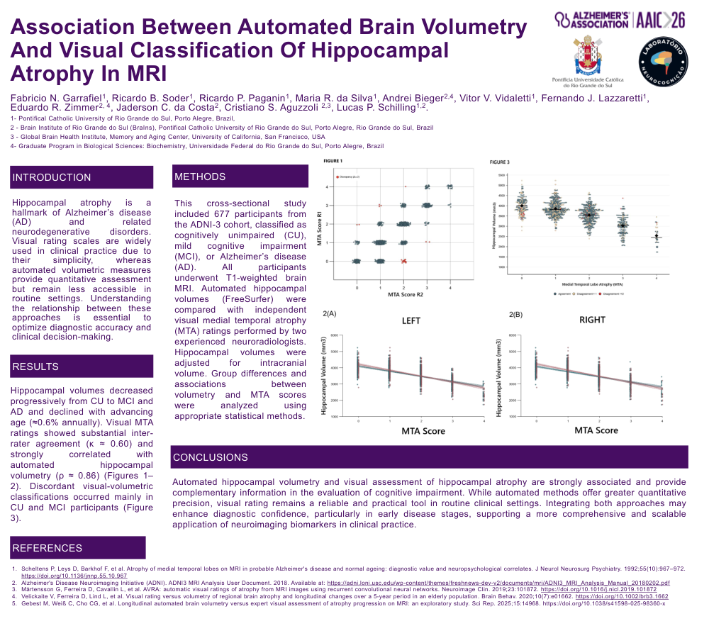
METHODS: This cross-sectional study included 677 participants from the ADNI-3 cohort, classified as cognitively unimpaired (CU), mild cognitive impairment (MCI), or Alzheimer’s disease (AD). All participants underwent T1-weighted brain MRI. Automated hippocampal volumes (FreeSurfer) were compared with independent visual medial temporal atrophy (MTA) ratings performed by two experienced neuroradiologists. Hippocampal volumes were adjusted for intracranial volume. Group differences and associations between volumetry and MTA scores were analyzed using appropriate statistical methods.
RESULTS: Hippocampal volumes decreased progressively from CU to MCI and AD and declined with advancing age (≈0.6% annually). Visual MTA ratings showed substantial inter-rater agreement (κ ≈ 0.60) and strongly correlated with automated hippocampal volumetry (ρ ≈ 0.86) (Figures 1–2). Discordant visual-volumetric classifications occurred mainly in CU and MCI participants (Figure 3).
CONCLUSION: Automated hippocampal volumetry and visual assessment of hippocampal atrophy are strongly associated and provide complementary information in the evaluation of cognitive impairment. While automated methods offer greater quantitative precision, visual rating remains a reliable and practical tool in routine clinical settings. Integrating both approaches may enhance diagnostic confidence, particularly in early disease stages, supporting a more comprehensive and scalable application of neuroimaging biomarkers in clinical practice.
Comments
Email is required but is not shown publicly.
Loading comments...