4,068 posters, 219 videos, 101 topics, 69 sessions, 14,365 authors, 6,128 institutions
ePostersLive by SciGen Technologies S.A. All rights reserved.
Alzheimer's Association International Conference 2026
July 12-15, 2026 | London, United Kingdom, and Online

2404
Macular Retinal Layer Thickness in Early Parkinson’s Disease
Poster Presenter
Part of Topic
Macular Retinal Layer Thickness in Early Parkinson’s Disease
Michael Zhu BA, Sejal Patel BS, Nikki Daniels BA, John Hsu BA, Rachel D’Cunha BS, Alex Choi BS, Alice Haystead BS,
Stephanie Chiu PhD, Burton Scott MD PhD, Dilraj S. Grewal MD FASRS, Sharon Fekrat MD FASRS
Departments of Ophthalmology and Neurology, Duke University School of Medicine
Background.
As a direct extension of the central nervous system, the retina frequently mirrors neurodegenerative processes in the brain.1-3
Optical Coherence Tomography (OCT) is a critical noninvasive tool for quantifying retinal microstructure.4
While prior OCT studies have established that Parkinson’s disease (PD) is associated with thinning in the inner retinal layers and photoreceptor layer, data regarding structural changes in the other layers remain incompletely described.5-7
Characterizing the structural changes across retinal layers could lead to the discovery of reliable, noninvasive biomarkers that enable earlier disease detection and objective monitoring.8,9
Purpose.
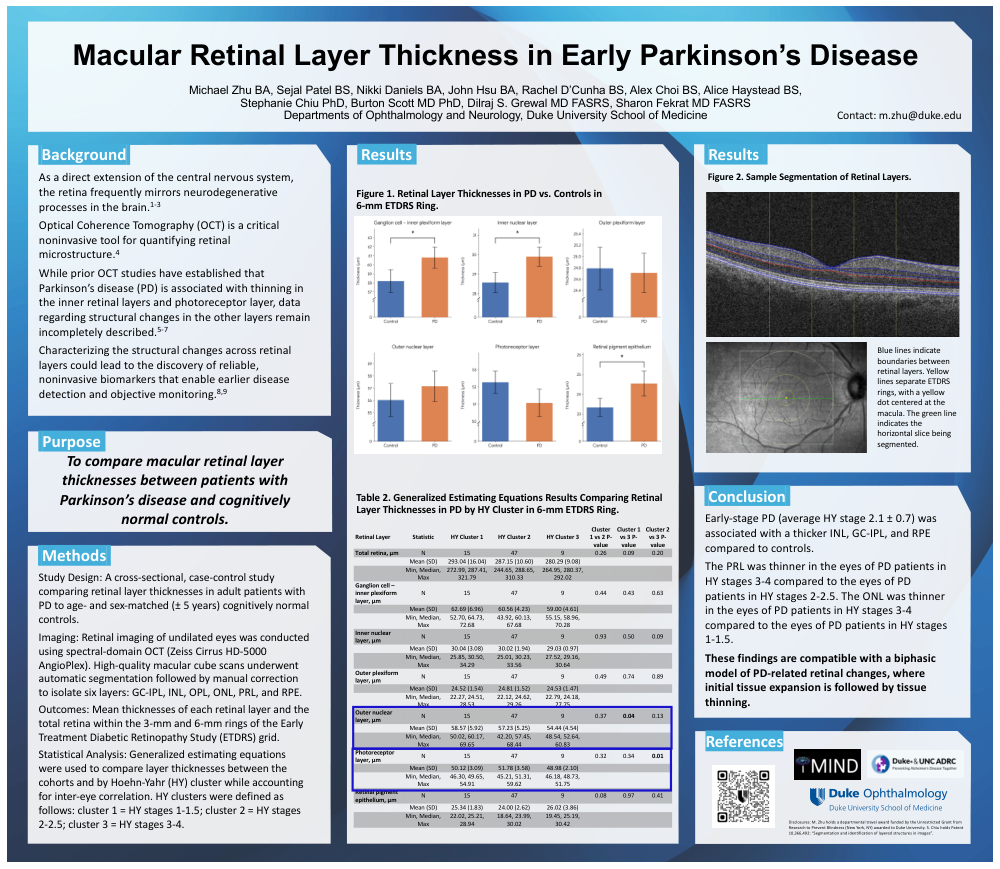
To compare macular retinal layer thicknesses between patients with Parkinson’s disease and cognitively normal controls.
Methods.
Study Design: A cross-sectional, case-control study comparing retinal layer thicknesses in adult patients with PD to age- and sex-matched (± 5 years) cognitively normal controls.
Imaging: Retinal imaging of undilated eyes was conducted using spectral-domain OCT (Zeiss Cirrus HD-5000 AngioPlex). High-quality macular cube scans underwent automatic segmentation followed by manual correction to isolate six layers: GC-IPL, INL, OPL, ONL, PRL, and RPE.
Outcomes: Mean thicknesses of each retinal layer and the total retina within the 3-mm and 6-mm rings of the Early Treatment Diabetic Retinopathy Study (ETDRS) grid.
Statistical Analysis: Generalized estimating equations were used to compare layer thicknesses between the cohorts and by Hoehn-Yahr (HY) cluster while accounting for inter-eye correlation. HY clusters were defined as follows: cluster 1 = HY stages 1-1.5; cluster 2 = HY stages 2-2.5; cluster 3 = HY stages 3-4.
Conclusion.
Early-stage PD (average HY stage 2.1 ± 0.7) was associated with a thicker INL, GC-IPL, and RPE compared to controls.
The PRL was thinner in the eyes of PD patients in HY stages 3-4 compared to the eyes of PD patients in HY stages 2-2.5. The ONL was thinner in the eyes of PD patients in HY stages 3-4 compared to the eyes of PD patients in HY stages 1-1.5.
These findings are compatible with a biphasic model of PD-related retinal changes, where initial tissue expansion is followed by tissue thinning.
Comments
Email is required but is not shown publicly.
Loading comments...