4,068 posters, 219 videos, 101 topics, 69 sessions, 14,365 authors, 6,128 institutions
ePostersLive by SciGen Technologies S.A. All rights reserved.
Alzheimer's Association International Conference 2026
July 12-15, 2026 | London, United Kingdom, and Online

3217
Modeling Network Tau Diffusion
Part of Topic
INTRODUCTION
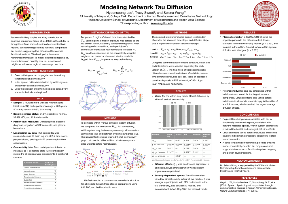
Tau neurofibrillary tangles are a key contributor to cognitive impairment (Vogel et al., 2020). Although tau is thought to diffuse across functionally connected brain regions, connected regions may not show comparable tau burden, suggesting that diffusion differs across functional systems. We developed a three-level hierarchical framework to model longitudinal regional tau accumulation and quantify how tau in connected neighbors influences regional tau change over time.
RESEARCH QUESTIONS
- Does pathological tau propagate over time along functional brain connectivity?
- Is tau spread better characterized by within-system or between-system connectivity?
- Does the strength of network-mediated spread vary across individuals and regions?
DATA
- Sample: 219 Alzheimer's Disease Neuroimaging Initiative (ADNI) participants (mean age = 70.5 years, SD = 6.8; range = 55–87; 51% male)
- Baseline clinical status: 54.8% cognitively normal, 32.4% MCI, and 12.8% dementia
- Person-level measures: Demographics, baseline diagnosis, cognition, APOE ε4 counts, and plasma biomarkers
- Longitudinal tau data: PET-derived tau was measured across 68 brain regions at 2–7 time points per participant, yielding 44,472 person-region-time observations.
- Connectivity data: Each participant contributed an individual 68 × 68 resting-state fMRI connectivity matrix; the 68 regions were grouped into 8 functional systems.
NETWORK DIFFUSION OF TAU
For person i, region r’s tau at time t was denoted by Y_tri . Each region’s diffusion exposure was defined as the tau burden in its functionally connected neighbors. After removing self-connections, each participant’s connectivity matrix was row-normalized to obtain W_i. D_tri was then calculated as the connectivity-weighted neighbor tau burden and entered into the model in lagged form D_t-1,ri to preserve temporal ordering.
METHODS
To compare within- versus between-system diffusion, we compared five versions of D_t-1: full connectivity, within-system only, between-system only, within-system upweighted (×2), and between-system upweighted (×2). The upweighted versions retained the full connectivity graph but doubled either within- or between-system edge weights before normalization. We first selected a common random-effects structure for all models through three staged comparisons using AIC, BIC, and likelihood-ratio tests.
The selected structure included person-level random effects for the intercept, time slope, and diffusion slope, plus a region-within-person random intercept:
Using this common random-effects structure, covariates and interactions were selected separately for each version of D_t-1. The final fixed-effects specifications differed across operationalizations. Candidate person-level covariates included age, sex, years of education, baseline diagnosis, APOE e4 count, ADAS-13, p-tau217/Aβ40, and Aβ42/Aβ40.
RESULTS
- Model fit: The within-only model fit best, followed by within×2 and full connectivity.
- Diffusion effect: D_t-1 was positive and significant in all models. It was strongest when within-system edges were emphasized.
- Severity-dependent spread: The diffusion effect varied by clinical severity in four of five models. It was stronger in participants with MCI or dementia in the full, within-only, and between×2 models, and increased with ADAS-Cog 13 in the within×2 model.
- Plasma biomarker: p-tau217/Aβ40 showed the opposite pattern to the diffusion effect. It was strongest in the between-only models (b ≈ 0.137) and weakest in the within×2 model, where within-system diffusion was strongest (b = 0.031).
- Heterogeneity: Regional tau differences within individuals accounted for the largest variance component. Diffusion effects also varied across individuals in all models, most strongly in the within×2 and full models, which also had the largest average diffusion effects.
CONCLUSIONS
- Regional tau change was associated with tau in functionally connected regions, consistent with network-mediated spread. Within-system connectivity provided the best fit and strongest diffusion effects.
- Diffusion effects varied across individuals and clinical severity, indicating heterogeneity in network-related tau accumulation.
- A three-level diffusion framework provides a way to model connectivity-coupled tau progression and supports future work on functional-system mapping and person-level predictors.
ACKNOWLEDGMENTS
Dr. Selena Wang is supported by the William H. Gates Sr. Fellowship from the Alzheimer’s Disease Data Initiative and P30AG072976.
REFERENCES
Vogel, J. W., Iturria-Medina, Y., Strandberg, O. T., et al. (2020). Spread of pathological tau proteins through communicating neurons in human Alzheimer's disease. Nature Communications, 11(1):2612.
Comments
Email is required but is not shown publicly.
Loading comments...