4,068 posters, 219 videos, 101 topics, 69 sessions, 14,365 authors, 6,128 institutions
ePostersLive by SciGen Technologies S.A. All rights reserved.
Alzheimer's Association International Conference 2026
July 12-15, 2026 | London, United Kingdom, and Online

3777
Microglia Expressing The Alzheimer's Disease Protective Variant Plcγ2-p522r Suppress The Nlrp3 Inflammasome And Mitochondrial Stress
Poster Presenter
Part of Topic
Background:
Objectives:
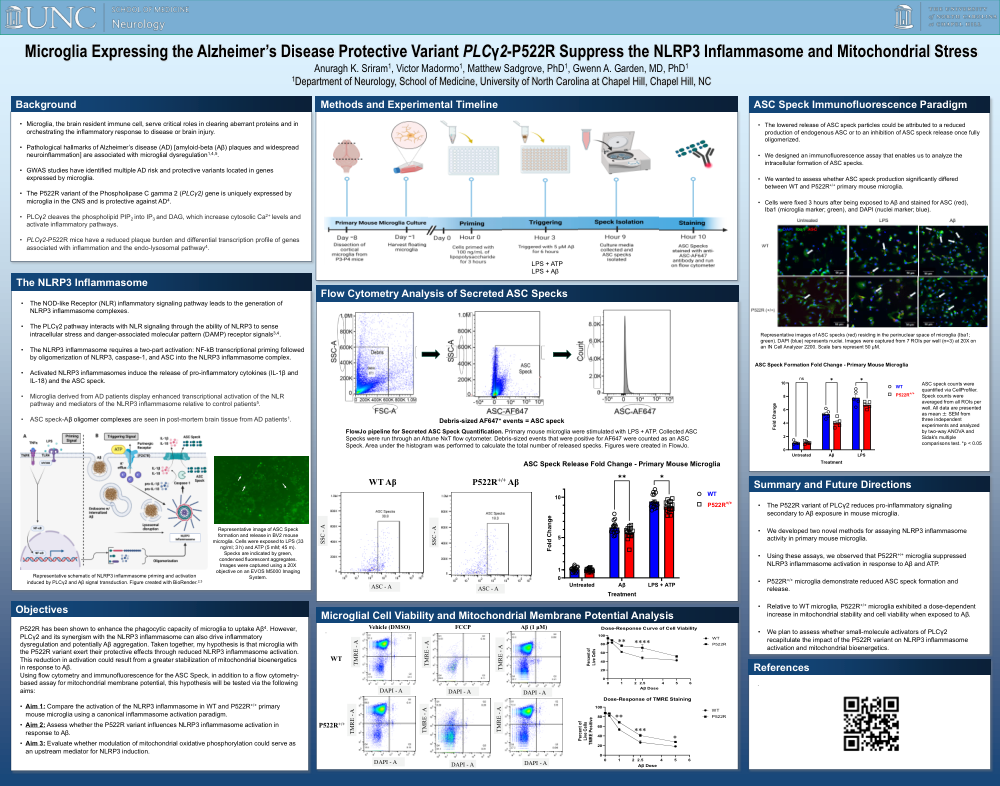
P522R has been shown to enhance the phagocytic capacity of microglia to uptake Aβ. However, PLCγ2 and its synergism with the NLRP3 inflammasome can also drive inflammatory dysregulation and potentially Aβ aggregation. Taken together, I hypothesize that microglia with the P522R variant exert their protective effects through reduced NLRP3 inflammasome activation. This reduction in activation could result from a greater stabilization of mitochondrial bioenergetics in response to Aβ.
Using flow cytometry and immunofluorescence for the ASC Speck, in addition to a flow cytometry-based assay for mitochondrial membrane potential, this hypothesis will be tested via the following aims:
Methods and Experimental Timeline:
Flow Cytometry Analysis of Secreted ASC Specks:
FlowJo pipeline for Secreted ASC Speck Quantification. Primary mouse microglia were stimulated with LPS + ATP. Collected ASC Specks were run through an Attune NxT flow cytometer. Debris-sized events that were positive for AF647 were counted as an ASC Speck. Area under the histogram was performed to calculate the total number of released specks. Figures were created in FlowJo.
Microglial Cell Viability and Mitochondrial Membrane Potential Analysis:
ASC Speck Immunofluorescence Paradigm:
• The lowered release of ASC speck particles could be attributed to a reduced production of endogenous ASC or to an inhibition of ASC speck release once fully oligomerized.
• We designed an immunofluorescence assay that enables us to analyze the intracellular formation of ASC specks.
• We wanted to assess whether ASC speck production significantly differed between WT and P522R+/+ primary mouse microglia.
• Cells were fixed 3 hours after being exposed to Aβ and stained for ASC (red), Iba1 (microglia marker; green), and DAPI (nuclei marker; blue).
ASC speck counts were quantified via CellProfiler. Speck counts were averaged from all ROIs per well. All data are presented as mean ± SEM from three independent experiments and analyzed by two-way ANOVA and Sidak's multiple comparisons test. *p < 0.05
Summary and Future Directions
• The P522R variant of PLCγ2 reduces pro-inflammatory signaling secondary to Aβ exposure in mouse microglia.
• We developed two novel methods for assaying NLRP3 inflammasome activity in primary mouse microglia.
• Using these assays, we observed that P522R+/+ microglia suppressed NLRP3 inflammasome activation in response to Aβ and ATP.
• P522R+/+ microglia demonstrate reduced ASC speck formation and release.
• Relative to WT microglia, P522R+/+ microglia exhibited a dose-dependent increase in mitochondrial stability and cell viability when exposed to Aβ.
• We plan to assess whether small-molecule activators of PLCγ2 recapitulate the impact of the P522R variant on NLRP3 inflammasome activation and mitochondrial bioenergetics.
Comments
Email is required but is not shown publicly.
Loading comments...