4,068 posters, 219 videos, 101 topics, 69 sessions, 14,365 authors, 6,128 institutions
ePostersLive by SciGen Technologies S.A. All rights reserved.
Alzheimer's Association International Conference 2026
July 12-15, 2026 | London, United Kingdom, and Online

9067
Postmortem, Dye-free Identification Of Potential RNA Granules In A Retina Of An Individual With FTLD-TDP
Poster Presenter
Authors
Melanie CW Campbell, Lyndsy Acheson, Erik L Mason, Ian R Mackenzie, Naomi C Futhey, Ging-Yuek Robin Hsiung, Veronica Hirsch-Reinshagen
Affiliations
University of British Columbia, Physics and Astronomy and School of Optometry and Vision Science University of Waterloo, Physics and Astronomy University of Waterloo
Part of Topic
Video
TAKE AWAY AND SUMMARY
THE FIRST POSTMORTEM, DYE-FREE IDENTIFICATION OF POTENTIAL RNA GRANULES IN A RETINA OF AN INDIVIDUAL WITH FTLD-TDP. IN VIVO DETECTION OF THESE GRANULES, WHICH MAY TRIGGER TDP-43 FORMATION INTO LARGER DEPOSITS, COULD BE AN EARLY DIAGNOSTIC
BACKGROUND AND INTRODUCTION
Previously, dye-free, with polarized light, we differentiated presumed TDP-43 deposits in 6 postmortem retinas from individuals with ALS and/or FTLD-TDP
from deposits in retinas of those with Alzheimer’s Disease. Here we describe additional small deposits, imaged in the retina with the largest number of
presumed TDP-43 deposits from an individual with FTLD-TDP. The small deposits appear consistent with RNA granules, including stress granules,
which have recently been suggested to trigger TDP-43 aggregation into fibrillar deposits. Retinal stress granules have only previously been reported
in association with light or chemical damage, optic nerve injury or ocular hypertension. This novel, patented method of retinal imaging in the living eye
could provide an inexpensive, dye-free, differential diagnostic that would be accessible to underserved populations. It would also enable appropriate,
early treatments and interventions.
METHODS
Eyes and brains were donated by 2 individuals with ALS, including 1 with concurrent FTLD-TDP, and 4 individuals with FTLD-TDP. Retinas were fixed
and flat-mounted and deposits were imaged using polarized light, dye-free. Here in only one of those retinas (of an individual with FTLD-TDP), we saw
and imaged, in polarized light, additional much smaller, approximately spherical deposits. We compared the sizes and strengths of the polarized
light signals of smaller with larger deposits.
RESULTS
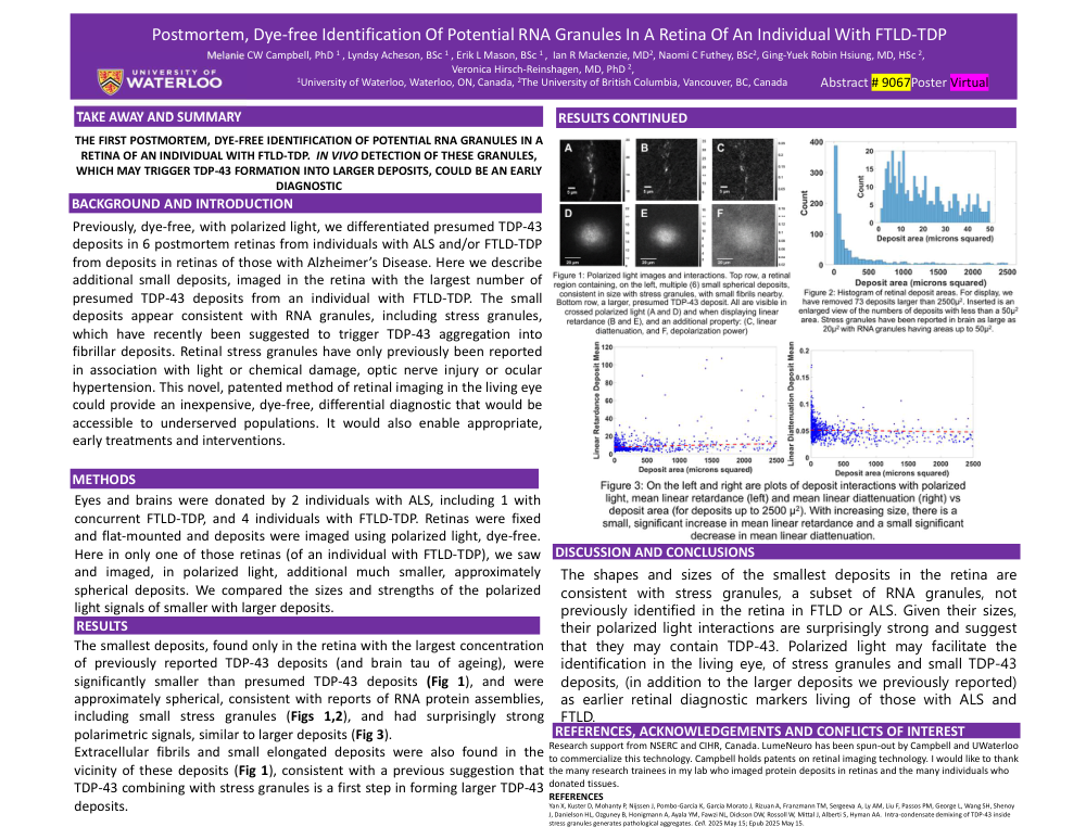
The smallest deposits, found only in the retina with the largest concentration of previously reported TDP-43 deposits (and brain tau of ageing), were
significantly smaller than presumed TDP-43 deposits (Fig 1), and were approximately spherical, consistent with reports of RNA protein assemblies,
including small stress granules (Figs 1,2), and had surprisingly strong polarimetric signals, similar to larger deposits (Fig 3). Extracellular fibrils and small elongated deposits were also found in the vicinity of these deposits (Fig 1), consistent with a previous suggestion that TDP-43 combining with stress granules is a first step in forming larger TDP-43 deposits.
DISCUSSION AND CONCLUSIONS
The shapes and sizes of the smallest deposits in the retina are consistent with stress granules, a subset of RNA granules, not previously identified in the retina in FTLD or ALS. Given their sizes, their polarized light interactions are surprisingly strong and suggest that they may contain TDP-43. Polarized light may facilitate the identification in the living eye, of stress granules and small TDP-43 deposits, (in addition to the larger deposits we previously reported) as earlier retinal diagnostic markers living of those with ALS and FTLD.
PLUS FIGURES AND CAPTIONS.
Comments
Email is required but is not shown publicly.
Loading comments...