4,068 posters, 219 videos, 101 topics, 69 sessions, 14,365 authors, 6,128 institutions
ePostersLive by SciGen Technologies S.A. All rights reserved.
Alzheimer's Association International Conference 2026
July 12-15, 2026 | London, United Kingdom, and Online

4232
Amyloid-vulnerable Cortical Regions Exhibit Higher Baseline Perfusion In A Subjective Cognitive Decline Cohort.
Poster Presenter
Authors
Lorenzo Gualco, Beatriz Padrela, Luigi Lorenzini, Leonard Pieperhoff, Mathijs B. J. Dijsselhof, Lyduine E. Collij, Ivan Gallesio, David Vàllez García, Marta Marquié, Mercè Boada, Juan Pablo Tartari, Oscar Sotolongo, Lluis Tárraga, Frederik Barkhof, Luca Roccatagliata, Matteo Pardini, Jan Petr, Henk J.M.M. Mutsaerts
Affiliations
null,
Part of Topic
Video
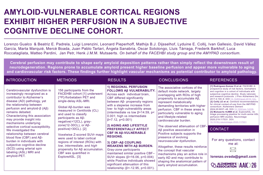
AMYLOID-VULNERABLE CORTICAL REGIONS EXHIBIT HIGHER PERFUSION IN A SUBJECTIVE COGNITIVE DECLINE COHORT.
Lorenzo Gualco & Beatriz E. Padrela, Luigi Lorenzini, Leonard Pieperhoff, Mathijs B.J. Dijsselhof, Lyduine E. Collij, Ivan Gallesio, David Vàllez García, Marta Marquié, Mercè Boada, Juan Pablo Tartari, Ángela Sanabria, Oscar Sotolongo, Lluis Tárraga, Frederik Barkhof, Luca Roccatagliata, Matteo Pardini, Jan Petr, Henk J.M.M. Mutsaerts. On behalf of the FACEHBI study group and the AMYPAD consortium.
Cerebral perfusion may contribute to shape early amyloid deposition patterns rather than simply reflect the downstream result of
neurodegeneration. Regions prone to accumulate amyloid present higher baseline perfusion and appear more vulnerable to aging
and cardiovascular risk factors. These findings further highlight vascular mechanisms as potential contributor to amyloid pathology.
INTRODUCTION
Cerebrovascular dysfunction is increasingly recognized as a contributor to Alzheimer’s disease (AD) pathology, yet the relationship between perfusion and amyloid-β (Aβ) remains debated. Characterizing this association may provide insight into mechanisms underlying selective cortical susceptibility. We investigated the relationship between cerebral blood flow (CBF) and Aβ-burden in individuals with subjective cognitive decline (SCD) using arterial spin labeling (ASL) MRI and amyloid-PET.
METHODS
156 participants from the FACEHBI cohort [1] underwent [18F]-florbetaben PET and single-delay ASL-MRI.
Global Aβ-burden was measured in Centiloids (CL) and used to classify participants as Aβ-negative(<12CL), gray-zone(12-30CL), or Aβ-positive(>30CL). [2]
Voxelwise Z-scored SUVr maps were used to label cortical regions of interest (ROIs) with low, intermediate, and high propensity for Aβ accumulation. CBF was quantified in ExploreASL. [3]
RESULTS
1) Regional perfusion follows Aβ vulnerability. Across each individual brain, CBF differed significantly between Aβ- propensity regions with a stepwise increase from low- to high-vulnerability areas (intermediate vs low β=14.59, p< 0.001; high vs intermediate β=7.12, p<0.001)
2) AGING and lifestyle preferentially affect CBF in Aβ-VULNERABLE REGIONS.
3) CBF–Aβ COUPLING WEAKENS WITH Aβ BURDEN. Gray-zone participants maintained similar positive CBF–SUVr slopes (β=16.06, p=0.004) while Positive individuals showed significant attenuation of this relationship (β=-12.95, p=0.001).
CONCLUSIONS
The associative cortices of the default mode network, largely overlapping with ROIs of high propensity to accumulate Aβ, represent metabolically demanding territories with higher perfusion. CBF in these areas is particularly vulnerable to aging and lifestyle-related cardiovascular burden.
The observed attenuation of CBF-Aβ positive association in Positive subjects supports the presence of evolving neurovascular dysfunction.
Altogether, these results reinforce the concept that vascular mechanisms play an active role in early AD and may contribute to shaping the anatomical pattern of early amyloid accumulation.
REFERENCES
[1] Rodriguez-Gomez O et al. FACEHBI: A prospective study of risk factors, biomarkers and cognition in a cohort of individuals with subjective cognitive decline. Study rationale and research protocols. J Prev Alzheimers Dis 2017;4:100–8. DOI:10.14283/jpad.2016.122.
[2] Collij LE et al. Centiloid recommendations for clinical context-of-use from the AMYPAD consortium. Alzheimers Dement 2024;20:9037–48. DOI:10.1002/alz.14336.
[3] Mutsaerts HJMM et al. ExploreASL: An image processing pipeline for multi-center ASL perfusion MRI studies. Neuroimage 2020;219:117031. DOI: 10.1016/j.neuroimage.2020.117031
CONTACT
For any questions, contact me at: lorenzo.ovada@gmail.com
Comments
Email is required but is not shown publicly.
Loading comments...