4,068 posters, 219 videos, 101 topics, 69 sessions, 14,365 authors, 6,128 institutions
ePostersLive by SciGen Technologies S.A. All rights reserved.
Alzheimer's Association International Conference 2026
July 12-15, 2026 | London, United Kingdom, and Online

5367
From Amyloid-related Imaging Abnormalities To Autopsy: Postmortem 7t Mri Following Anti-amyloid Immunotherapy In Alzheimer’S Disease
Poster Presenter
Part of Topic
Introduction:
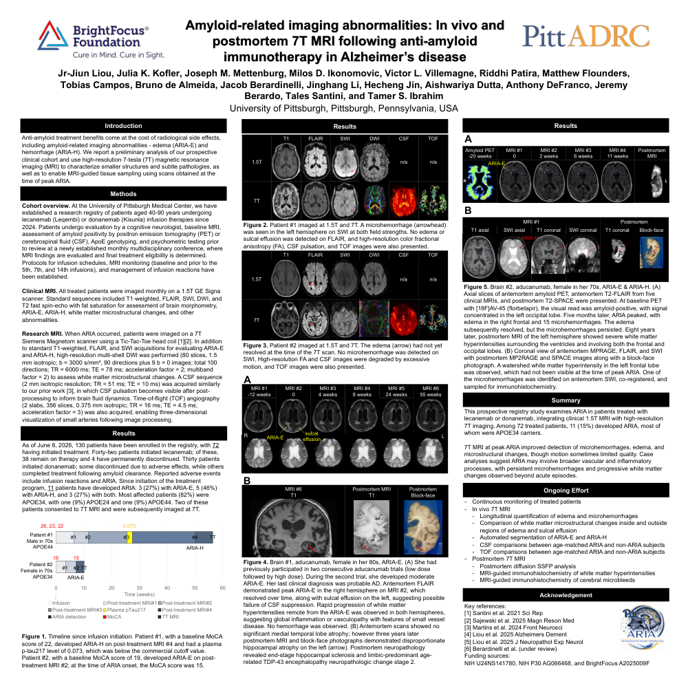
Anti-amyloid treatment benefits come at the cost of radiological side effects, including amyloid-related imaging abnormalities - edema (ARIA-E) and hemorrhage (ARIA-H). We report a preliminary analysis of our prospective clinical cohort and use high-resolution 7-tesla (7T) magnetic resonance imaging (MRI) to characterize smaller structures and subtle pathologies, as well as to enable MRI-guided tissue sampling using scans obtained at the time of peak ARIA.
Methods:
Cohort overview. At the University of Pittsburgh Medical Center, we have established a research registry of patients aged 40-90 years undergoing lecanemab (Leqembi) or donanemab (Kisunla) infusion therapies since 2024. Patients undergo evaluation by a cognitive neurologist, baseline MRI, assessment of amyloid positivity by positron emission tomography (PET) or cerebrospinal fluid (CSF), ApoE genotyping, and psychometric testing prior to review at a newly established monthly multidisciplinary conference, where MRI findings are evaluated and final treatment eligibility is determined. Protocols for infusion schedules, MRI monitoring (baseline and prior to the 5th, 7th, and 14th infusions), and management of infusion reactions have been established.
Clinical MRI. All treated patients were imaged monthly on a 1.5T GE Signa scanner. Standard sequences included T1-weighted, FLAIR, SWI, DWI, and T2 fast spin-echo with fat saturation for assessment of brain morphometry, ARIA-E, ARIA-H, white matter microstructural changes, and other abnormalities.
Research MRI. When ARIA occurred, patients were imaged on a 7T Siemens Magnetom scanner using a Tic-Tac-Toe head coil [1][2]. In addition to standard T1-weighted, FLAIR, and SWI acquisitions for evaluating ARIA-E and ARIA-H, high-resolution multi-shell DWI was performed (80 slices, 1.5 mm isotropic; b = 3000 s/mm², 90 directions plus 9 b = 0 images; total 100 directions; TR = 6000 ms; TE = 78 ms; acceleration factor = 2; multiband factor = 2) to assess white matter microstructural changes. A CSF sequence (2 mm isotropic resolution; TR = 51 ms; TE = 10 ms) was acquired similarly to our prior work [3], in which CSF pulsation becomes visible after post-processing to inform brain fluid dynamics. Time-of-flight (TOF) angiography (2 slabs, 356 slices, 0.375 mm isotropic; TR = 16 ms; TE = 4.5 ms; acceleration factor = 3) was also acquired, enabling three-dimensional visualization of small arteries following image processing.
Results:
As of June 8, 2026, 130 patients have been enrolled in the registry, with 72 having initiated treatment. Forty-two patients initiated lecanemab; of these, 38 remain on therapy and 4 have permanently discontinued. Thirty patients initiated donanemab; some discontinued due to adverse effects, while others completed treatment following amyloid clearance. Reported adverse events include infusion reactions and ARIA. Since initiation of the treatment program, 11 patients have developed ARIA: 3 (27%) with ARIA-E, 5 (46%) with ARIA-H, and 3 (27%) with both. Most affected patients (82%) were APOE34, with one (9%) APOE24 and one (9%) APOE44. Two of these patients consented to 7T MRI and were subsequently imaged at 7T.
Summary:
This prospective registry study examines ARIA in patients treated with lecanemab or donanemab, integrating clinical 1.5T MRI with high-resolution 7T imaging. Among 72 treated patients, 11 (15%) developed ARIA, most of whom were APOE34 carriers. 7T MRI at peak ARIA improved detection of microhemorrhages, edema, and microstructural changes, though motion sometimes limited quality. Case analyses suggest ARIA may involve broader vascular and inflammatory processes, with persistent microhemorrhages and progressive white matter changes observed beyond acute episodes.
Comments
Email is required but is not shown publicly.
Loading comments...