4,068 posters, 219 videos, 101 topics, 69 sessions, 14,365 authors, 6,128 institutions
ePostersLive by SciGen Technologies S.A. All rights reserved.
Alzheimer's Association International Conference 2026
July 12-15, 2026 | London, United Kingdom, and Online

13743
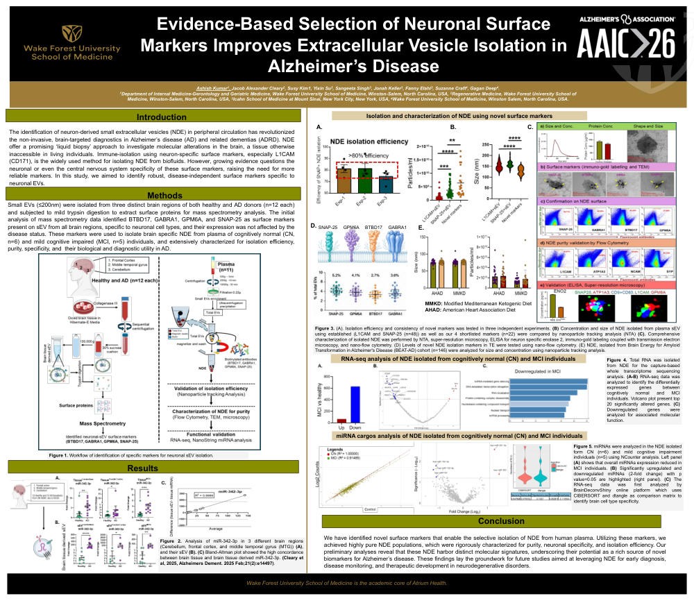
Evidence Based Selection Of Neuronal Surface Markers Improves Extracellular Vesicle Isolation In Alzheimer’S Disease
Poster Presenter
Authors
Ashish Kumar, Jacob Alexander Cleary, Susy Kim, Yixin Su, Sangeeta Singh, Jonah Keller, Fanny Elahi, Suzanne Ceaft, Gagan Deep
Affiliations
Department of Internal Medicine-Gerontology and Geriatric Medicine, Wake Forest University School of Medicine, Winston-Salem, North Carolina, USA, Icahn School of Medicine at Mount Sinai, New York City, New York, USA, Wake Forest University School of Medicine, Winston Salem, North Carolina, USA
Part of Topic
Biomarkers -> Biomarkers (non-neuroimaging) -> Fluid biomarkers
Abstract:
Background: Blood-based biomarker development for Alzheimer’s disease (AD) is limited by poor access to brain neuron-specific molecular information from the periphery. Most circulating analytes lack brain specificity and are dominated by peripheral signals, resulting in dilution of neuronal information. Neuron-derived small extracellular vesicles (NDE) offer a biologically relevant liquid biopsy approach by enriching neuron-associated signals within a protective lipid bilayer, improving their stability and detectability in blood. Although immuno-isolation using neuronal surface markers, particularly L1CAM, is widely used, growing evidence questions the specificity and consistency of current markers, underscoring the need for a systematic, evidence-based framework for NDE marker selection.
Methods: Small EVs were isolated from three brain regions (frontal cortex, middle temporal gyrus, and cerebellum) obtained from control and AD donors (n = 12 per group). EV surface proteins were selectively shaved using mild trypsin digestion and analyzed by mass spectrometry. Deconvolution analysis identified proteins consistently present on the brain tissue-derived EV surface, unaffected by disease status, and neuronally expressed. Identified markers were used to immuno-isolate NDE from plasma and characterization using nanoparticle tracking analysis, super-resolution microscopy, flow cytometry, and immunogold labeling. Transcriptomic profiling was performed using RNA sequencing and NanoString miRNA analysis.
Results: Surface proteomic profiling identified four candidate neuronal markers, SNAP-25, GPM6A, BTBD17, and GABRA1, consistently present on brain tissue-derived EVs across regions and disease states. Nano-flow cytometry confirmed their presence (2-6%) in circulating plasma total EVs. NDE isolated using these markers were enriched for neuronal and synaptic proteins and RNAs, with reduced contribution from non-neuronal vesicles. In plasma from amyloid β positive individuals, NDEs showed significant downregulation of both mRNA and miRNA compared with controls, consistent with transcriptional dysregulation and synaptic dysfunction in AD. These markers were further applied to isolate NDEs from participants (n = 146) enrolled in a ketogenic diet intervention trial, where NDE surface levels of monocarboxylate transporter-2 showed significant pre- versus post-intervention changes.
Conclusion: This study defines a tissue-informed framework for neuronal surface marker selection and identifies a novel, disease-independent marker panel for robust NDE isolation, supporting the use of NDEs for brain-specific molecular profiling and biomarker development in AD.
Comments
Email is required but is not shown publicly.
Loading comments...