4,068 posters, 219 videos, 101 topics, 69 sessions, 14,365 authors, 6,128 institutions
ePostersLive by SciGen Technologies S.A. All rights reserved.
Alzheimer's Association International Conference 2026
July 12-15, 2026 | London, United Kingdom, and Online

6268
Ultrashort Echo Time Magnetic Resonance Imaging Of Myelin And Iron In Parkinson's Disease
Part of Topic
Biomarkers -> Neuroimaging -> Case study
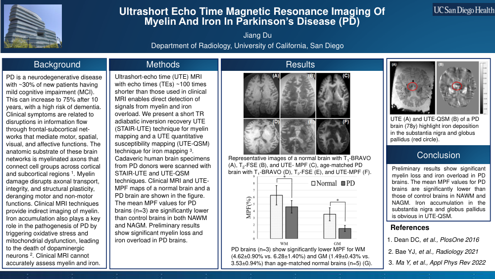
Parkinson’s disease (PD) is a common neurodegenerative disorder. About 30% of newly diagnosed PD patients have mild cognitive impairment (MCI), and this can increase to 75% after 10 years, with a high risk of progressing to dementia. Clinical symptoms are related to disruptions in information flow through frontal-subcortical networks that mediate motor, spatial, visual, and affective functions. The anatomic substrate of these brain networks is myelinated axons that connect cell groups across cortical and subcortical regions. Myelin damage disrupts axonal transport, integrity, and structural plasticity, deranging motor and non-motor functions. Clinical MRI techniques provide indirect imaging of myelin. They lack specificity for myelin damage, especially for partial demyelination in lesions and in normal-appearing white matter (NAWM) and gray matter (NAGM). Iron accumulation also plays a key role in the pathogenesis of PD by triggering oxidative stress and mitochondrial dysfunction, leading to the death of dopaminergic neurons. Clinical MRI cannot accurately assess myelin and iron content. Direct myelin imaging and quantitative iron mapping may provide a more specific diagnosis of PD. However, myelin has an ultrashort T2 (<<1 ms)and isinvisible with clinical MRI. Iron overload also tends to reduce T2* and is difficult to quantify accurately with clinical MRI. It is crucial to develop novel MRI techniques that can map myelin and iron in lesions and in NAWM and NAGM, enabling more specific diagnosis and treatment monitoring of PD. 3D ultrashort-echo time (UTE) sequences with echo times (TEs) ~100 times shorter than those used in clinical MRI enable direct detection of signals from myelin and iron overload. In this abstract, we present a short TR adiabatic inversion recovery UTE (STAIR-UTE) technique for myelin mapping and a UTE quantitative susceptibility mapping (UTE-QSM) technique for iron mapping. Cadaveric human brain specimens from PD donors were scanned with STAIR-UTE and UTE-QSM techniques. Clinical MRI and UTE-MPF maps of a normal brain and a PD brain are shown in the figure. The mean MPF values for PD brains (n=3) are significantly lower than control brains in both NAWM and NAGM. Preliminary results show significant myelin loss and iron overload in PD brains.
Comments
Email is required but is not shown publicly.
Loading comments...