4,068 posters, 219 videos, 101 topics, 69 sessions, 14,365 authors, 6,128 institutions
ePostersLive by SciGen Technologies S.A. All rights reserved.
Alzheimer's Association International Conference 2026
July 12-15, 2026 | London, United Kingdom, and Online

2930
Retinal Accumulation Of α-synuclein After Intracranial Preformed Fibril Injection
Poster Presenter
Authors
Teo Sällberg, Valentina Cesaroni, Jessica Santiago, Angela Censi-Nilsson, Malin Wennström
Affiliations
Clinical Memory Research Unit, Department of Clinical Sciences Malmö, Lund University, Malmö, Sweden., Diabetes and Brain Function Unit, Department of Experimental Medical Science, Lund University, Lund, Sweden, Basal Ganglia Pathophysiology Unit, Department of Experimental Medical Science, Lund University, Lund, Sweden
Part of Topic
α‑Synuclein-Induced Increase in DOPA Decarboxylase-Expressing Cells in the Retina
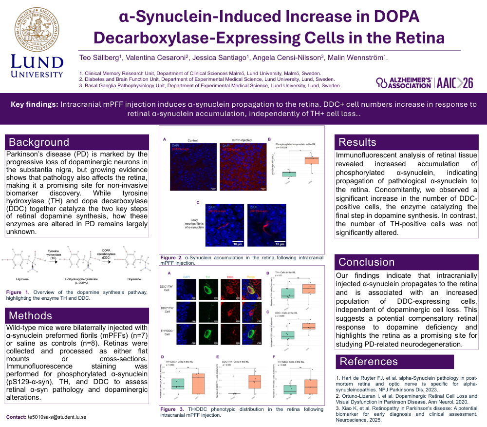
Key findings: Intracranial mPFF injection induces α-synuclein propagation to the retina. DDC+ cell numbers increase in response to retinal α-synuclein accumulation, independently of TH+ cell loss..
Background:
Parkinson’s disease (PD) is marked by the progressive loss of dopaminergic neurons in the substantia nigra, but growing evidence shows that pathology also affects the retina, making it a promising site for non‑invasive biomarker discovery. While tyrosine hydroxylase (TH) and dopa decarboxylase (DDC) together catalyze the two key steps of retinal dopamine synthesis, how these enzymes are altered in PD remains largely unknown.
Methods:
Wild‑type mice were bilaterrally injected with α‑synuclein preformed fibrils (mPFFs) (n=7) or saline as controls (n=8). Retinas were collected and processed as either flat mounts or cross‑sections. Immunofluorescence staining was performed for phosphorylated α‑synuclein (pS129‑α‑syn), TH, and DDC to assess retinal α‑syn pathology and dopaminergic alterations.
Results:
Immunofluorescent analysis of retinal tissue revealed increased accumulation of phosphorylated α‑synuclein, indicating propagation of pathological α‑synuclein to the retina. Concomitantly, we observed a significant increase in the number of DDC-positive cells, the enzyme catalyzing the final step in dopamine synthesis. In contrast, the number of TH-positive cells was not significantly altered.
Conclusion:
Our findings indicate that intracranially injected α‑synuclein propagates to the retina and is associated with an increased population of DDC‑expressing cells, independent of dopaminergic cell loss. This suggests a potential compensatory retinal response to dopamine deficiency and highlights the retina as a promising site for studying PD‑related neurodegeneration.
Comments
Email is required but is not shown publicly.
Loading comments...