4,068 posters, 219 videos, 101 topics, 69 sessions, 14,365 authors, 6,128 institutions
ePostersLive by SciGen Technologies S.A. All rights reserved.
Alzheimer's Association International Conference 2026
July 12-15, 2026 | London, United Kingdom, and Online

8651
Brain Networks In Motion: EEG Signatures Distinguishing Amnestic And Non-amnestic MCI In A Visual Short-term Memory Binding Task
Poster Presenter
Authors
Melody N. Sequeira, Avalon Campbell-Cousins, Rodrigo A Gonzalez-Montealegre, Alfredis Gonzalez-Hernandez, Jasmin Bonilla-Santos, Mario Parra, Javier Escudero
Affiliations
Institute of Neuroscience and Cardiovascular Research, College of Medicine and Veterinary Medicine, University of Edinburgh, UK, IDCOM, School of Engineering, University of Edinburgh, UK, Surcolombiana University, Neiva, Huila, Colombia, Cooperative University of Colombia, Psychology, University of Strathclyde, UK
Part of Topic
Brain Networks in Motion: EEG Signatures Distinguishing Amnestic and Non-Amnestic MCI in a Visual Short-Term Memory Binding Task.
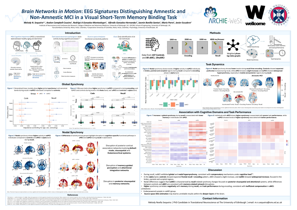
Mild cognitive impairment (MCI) is a transitional state between healthy aging and dementia, encompassing subtypes like amnestic (aMCI) and non-amnestic MCI (naMCI). While aMCI often precedes Alzheimer’s disease, naMCI may progress to other dementias. Task-based Electroencephalography (EEG) offers a cost-effective, non-invasive means to measure brain activity during cognitive processes. Here, we analysed EEG source-space signals from individuals with aMCI, naMCI and healthy controls to uncover functional connectivity patterns that could aid in early MCI subtype differentiation.
Figure 1: Generalized linear models adjusting for age, sex and schooling showhigher global synchrony in all canonical bands during recall in naMCIindividuals compared to controls.
Figure 2: Wilcoxon tests show higher synchrony in naMCI compared to during encoding, and naMCI and controls during recall in the theta band, and aMCI and controls in alpha band.
Figure 3: Nodal synchrony shows higher activity in naMCI individuals compared to controls and aMCI in alpha band during recall.
Figure 4: Difference of activity across groups highlight disruption in cognition-specific functional pathways in aMCI and naMCI during recall in alpha band.
- Disruption of posterior cortical association networks involving default mode, visuospatial and thalamocortical systems.
- Disruption of memory-guided perception and attentional integration networks.
- Disruption in posterior visuospatial and memory networks.
Figure 5: Nodal synchrony shows trends of higher activity in naMCIindividuals in limbic, parietal and occipital regions during recall compared to encoding phase in alpha band.
Figure 6: Nodal synchrony showed lower trends during recall than encoding. Controls showed expected prefrontal increase during recall, while aMCI showed a slight increase and naMCI showed widespread hypersynchrony, especially in medial and posterior regions during recall.
Figure 7: Increase in global synchrony during recall is associated with lowermemory in individuals with aMCI.
Figure 8: Individuals with aMCI shows higher synchrony is associated with poorer task performance, while naMCI showed trends of higher synchronyassociated with better performance.
Discussion:
Limitations:
Comments
Email is required but is not shown publicly.
Loading comments...