4,068 posters, 219 videos, 101 topics, 69 sessions, 14,365 authors, 6,128 institutions
ePostersLive by SciGen Technologies S.A. All rights reserved.
Alzheimer's Association International Conference 2026
July 12-15, 2026 | London, United Kingdom, and Online

10071
Effects Of Lecanemab On aß And Tau Pathology And Biomarkers In A Knock-in Mouse Model Of Alzheimer's Disease
Poster Presenter
Authors
Nils Lindblom, Niklas Mattson-Carlgren, Haoyang Zhang, Nicola Fattorelli, Tim Meese, Lena Jutz, Anne Wienand, Anika Perdok, Lluís Camprubí-Ferrer, Megg Garcia Ryde, Rosalía Fernández-Calle, Linda Söderberg, Christer Möller, Tomas Deierborg, Renzo Mancuso, Gunnar Gouras, Oskar Hansson
Affiliations
1. Clinical Memory Research Unit, Lund University, Lund, Sweden, 2 Department of Experimental Medical Science, Experimental Dementia Research Unit, Lund University, 1. Clinical Memory Research Unit, Lund University, Lund, Sweden 3. Memory Clinic, Skåne University Hospital, Malmö, Sweden, Clinical Memory Research Unit, Lund University, Lund, Sweden, 4 Microglia and Inflammation in Neurological Disorders (MIND) Lab, VIB Center for Molecular Neurology, VIB, Antwerp, Belgium, 5 Department of Biomedical Sciences, University of Antwerp, Antwerp, Belgium, Department of Experimental Medical Science, Experimental Neuroinflammation Laboratory, Lund University, Lund, Sweden, Department of Experimental Medical Science, Experimental Dementia Research Unit, Lund University, Lund, Sweden, Bioarctic AB 11251 Stockholm, Sweden
Part of Topic
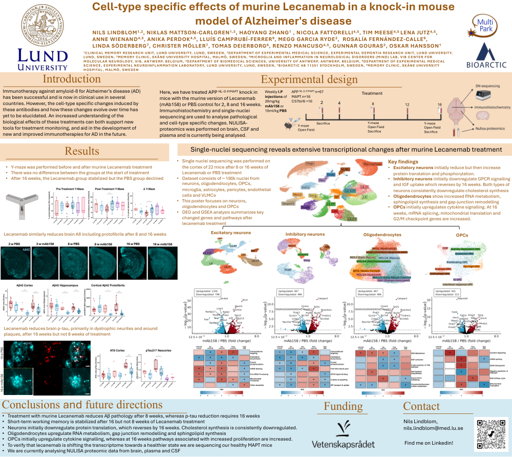
Cell-type specific effects of murine Lecanemab in a knock-in mouse model of Alzheimer's disease
Introduction
Immunotherapy against amyloid-ß for Alzheimer’s disease (AD) has been successful and is now in clinical use in several countries. However, the cell-type specific changes induced by these antibodies and how these changes evolve over time has yet to be elucidated. An increased understanding of the biological effects of these treatments can both support new tools for treatment monitoring, and aid in the development of new and improved immunotherapies for AD in the future.
Experimental Design
Here, we have treated APP NL-G-F/MAPT knock in mice with the murine version of Lecanemab (mAb158) or PBS control for 2, 8 and 16 weeks.Confirm and proceed »
Immunohistochemistry and single-nuclei sequencing are used to analyse pathological and cell-type specific changes. NULISA-proteomics was performed on brain, CSF and plasma and is currently being analysed.
Results
After 16 weeks, the Lecanemab group stabilized but the PBS group declined
Lecanemab similarly reduces brain Aß including protofibrils after 8 and 16 weeks
Lecanemab reduces brain p-tau, primarily in dystrophic neurites and around plaques, after 16 weeks but not 8 weeks of treatment
Single-nuclei sequencing reveals extensive transcriptional changes after murine Lecanemab treatment
Key findings
Comments
Email is required but is not shown publicly.
Loading comments...