4,068 posters, 219 videos, 101 topics, 69 sessions, 14,365 authors, 6,128 institutions
ePostersLive by SciGen Technologies S.A. All rights reserved.
Alzheimer's Association International Conference 2026
July 12-15, 2026 | London, United Kingdom, and Online

6352
Investigating The Relationship Between Endo-lysosomal Dysfunction And TDP-43 Pathology In Alzheimer’s Disease
Poster Presenter
Authors
Jordan Ogg, Mamatha Damodarasmy, Chizuru Kinoshita, Martin Darvas, Caitlin Latimer, Jessica Young
Affiliations
Part of Topic
Investigating The Relationship Between Endo-lysosomal Dysfunction And TDP-43 Pathology In Alzheimer’s Disease
Jordan Danielle Ogg, Mamatha Damodarasamy, Chizuru Kinoshita, Martin Darvas, PhD, Caitlin Latimer, Jessica Young.
Department of Laboratory Medicine and Pathology, University of Washington, Seattle, WA, USA.
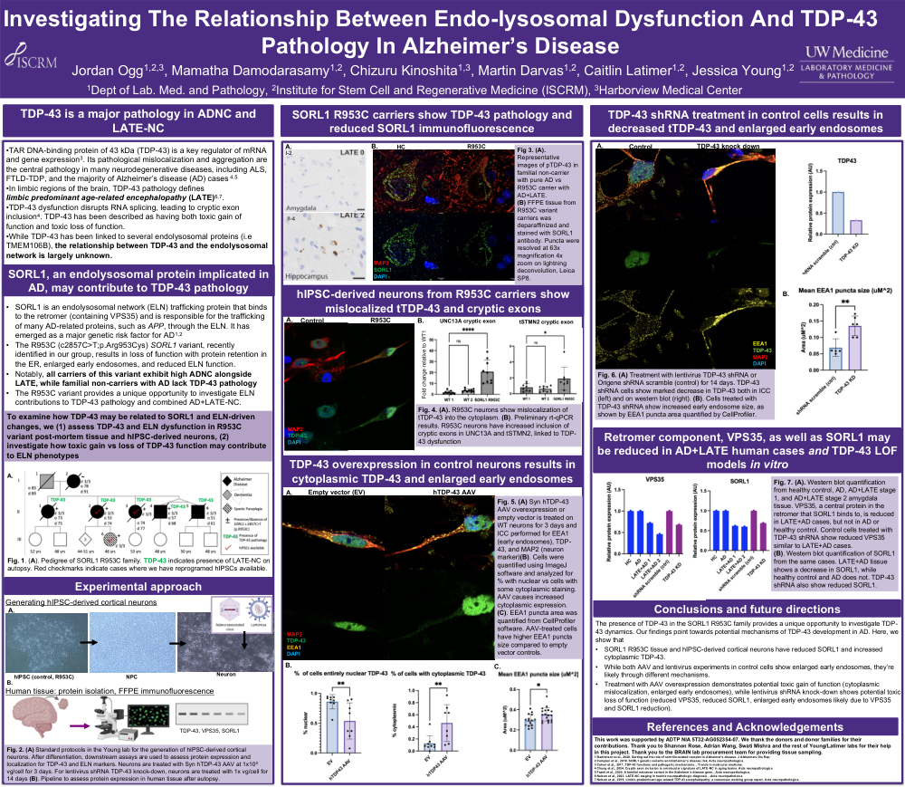
TDP-43 is a major pathology in ADNC and LATE-NC
SORL1, an endolysosomal protein implicated in AD, may contribute to TDP-43 pathology
To examine how TDP-43 may be related to SORL1 and ELN-driven changes, we (1) assess TDP-43 and ELN dysfunction in R953C variant post-mortem tissue and hIPSC-derived neurons, (2) investigate how toxic gain vs loss of TDP-43 function may contribute to ELN phenotypes
Methods: generating hiPSC-derived cortical neurons, human tissue protein isolation and FFPE immunofluorescence
Standard protocols in the Young lab for the generation of hIPSC-derived cortical neurons. After differentiation, downstream assays are used to assess protein expression and localization for TDP-43 and ELN markers. Neurons are treated with Syn hTDP-43 AAV at 1x10⁴ vg/cell for 3 days. For lentivirus shRNA TDP-43 knock-down, neurons are treated with 1x vg/cell for 14 days (B). Pipeline to assess protein expression in human tissue after autopsy.
Results
SORL1 R953C carriers show TDP-43 pathology and reduced SORL1 immunofluorescence
hIPSC-derived neurons from R953C carriers show mislocalizedtTDP-43 and cryptic exons
TDP-43 overexpression in control neurons results in cytoplasmic TDP-43 and enlarged early endosomes
TDP-43 shRNA treatment in control cells results in decreased tTDP-43 and enlarged early endosomes
Retromer component, VPS35, as well as SORL1 may be reduced in AD+LATE human cases and TDP-43 LOF models in vitro
Conclusions and future directions
The presence of TDP-43 in the SORL1 R953C family provides a unique opportunity to investigate TDP-43 dynamics. Our findings point towards potential mechanisms of TDP-43 development in AD. Here, we show that
Comments
Email is required but is not shown publicly.
Loading comments...