4,068 posters, 219 videos, 101 topics, 69 sessions, 14,365 authors, 6,128 institutions
ePostersLive by SciGen Technologies S.A. All rights reserved.
Alzheimer's Association International Conference 2026
July 12-15, 2026 | London, United Kingdom, and Online

9437
7T MRI Virtual Histology Of Choroid Plexus In Aging And Alzheimer’s Disease
Poster Presenter
Authors
Part of Topic
7T MRI Virtual Histology of Choroid Plexus in Aging and Alzheimer’s Disease
Chenyang Li1,
Zhe Sun1,
Dominique Leitner2,
Li Jiang1,
Thomas Wisniewski2,
Arline Faustin2
Youssef Z Wadghiri1,
Jiangyang Zhang1,
Yulin Ge1
1 Department of Radiology, NYU Grossman School of Medicine, New York, NY
2 Department of Neurology, NYU Grossman School of Medicine, New York, NY
1. Introduction:
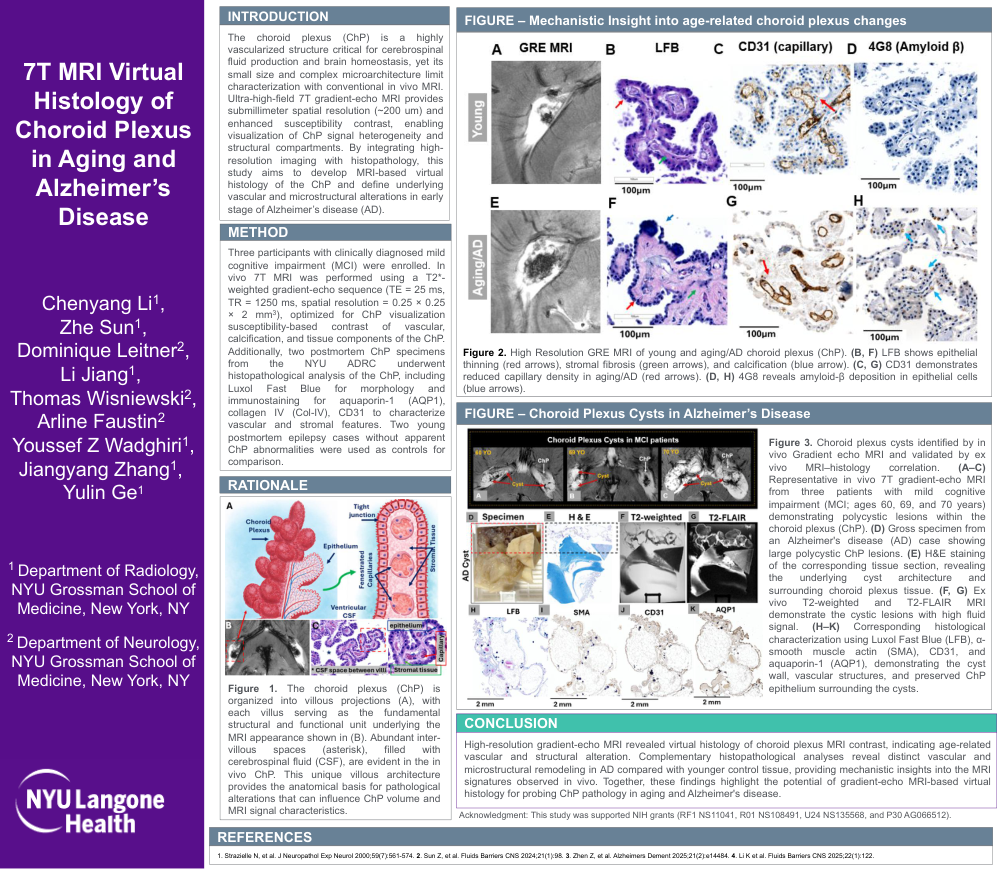
The choroid plexus (ChP) is a highly vascularized structure critical for cerebrospinal fluid production and brain homeostasis, yet its small size and complex microarchitecture limit characterization with conventional in vivo MRI. Ultra-high-field 7T gradient-echo MRI provides submillimeter spatial resolution (~200 um) and enhanced susceptibility contrast, enabling visualization of ChP signal heterogeneity and structural compartments. By integrating high-resolution imaging with histopathology, this study aims to develop MRI-based virtual histology of the ChP and define underlying vascular and microstructural alterations in early stage of Alzheimer’s disease (AD).
2. Method:
Three participants with clinically diagnosed mild cognitive impairment (MCI) were enrolled. In vivo 7T MRI was performed using a T2*-weighted gradient-echo sequence (TE = 25 ms, TR = 1250 ms, spatial resolution = 0.25 × 0.25 × 2 mm3), optimized for ChP visualization susceptibility-based contrast of vascular, calcification, and tissue components of the ChP. Additionally, two postmortem ChP specimens from the NYU ADRC underwent histopathological analysis of the ChP, including Luxol Fast Blue for morphology and immunostaining for aquaporin-1 (AQP1), collagen IV (Col-IV), CD31 to characterize vascular and stromal features. Two young postmortem epilepsy cases without apparent ChP abnormalities were used as controls for comparison.
3. Rationale:
Figure 1. The choroid plexus (ChP) is organized into villous projections (A), with each villus serving as the fundamental structural and functional unit underlying the MRI appearance shown in (B). Abundant inter-villous spaces (asterisk), filled with cerebrospinal fluid (CSF), are evident in the in vivo ChP. This unique villous architecture provides the anatomical basis for pathological alterations that can influence ChP volume and MRI signal characteristics.
4. FIGURE – Mechanistic Insight into age-related choroid plexus changes.
Figure 2. High Resolution GRE MRI of young and aging/AD choroid plexus (ChP). (B, F) LFB shows epithelial thinning (red arrows), stromal fibrosis (green arrows), and calcification (blue arrow). (C, G) CD31 demonstrates reduced capillary density in aging/AD (red arrows). (D, H) 4G8 reveals amyloid-β deposition in epithelial cells (blue arrows).
5. FIGURE – Choroid Plexus Cysts in Alzheimer’s Disease
Figure 3. Choroid plexus cysts identified by in vivo Gradient echo MRI and validated by ex vivo MRI–histology correlation. (A–C) Representative in vivo 7T gradient-echo MRI from three patients with mild cognitive impairment (MCI; ages 60, 69, and 70 years) demonstrating polycystic lesions within the choroid plexus (ChP). (D) Gross specimen from an Alzheimer's disease (AD) case showing large polycystic ChP lesions. (E) H&E staining of the corresponding tissue section, revealing the underlying cyst architecture and surrounding choroid plexus tissue. (F, G) Ex vivo T2-weighted and T2-FLAIR MRI demonstrate the cystic lesions with high fluid signal. (H–K) Corresponding histological characterization using Luxol Fast Blue (LFB), α-smooth muscle actin (SMA), CD31, and aquaporin-1 (AQP1), demonstrating the cyst wall, vascular structures, and preserved ChP epithelium surrounding the cysts.
6. Conclusion:
High-resolution gradient-echo MRI revealed virtual histology of choroid plexus MRI contrast, indicating age-related vascular and structural alteration. Complementary histopathological analyses reveal distinct vascular and microstructural remodeling in AD compared with younger control tissue, providing mechanistic insights into the MRI signatures observed in vivo. Together, these findings highlight the potential of gradient-echo MRI-based virtual histology for probing ChP pathology in aging and Alzheimer's disease.
Comments
Email is required but is not shown publicly.
Loading comments...