4,068 posters, 219 videos, 101 topics, 69 sessions, 14,365 authors, 6,128 institutions
ePostersLive by SciGen Technologies S.A. All rights reserved.
Alzheimer's Association International Conference 2026
July 12-15, 2026 | London, United Kingdom, and Online

10063
Cortical Microstructural MRI Changes Are Associated With CSF APP Across The Alzheimer’S Disease Clinical Continuum
Poster Presenter
Authors
Mario Torso, Gerard R. Ridgway, Pegah Khosropanah, Ian Hardingham, Steven A. Chance
Affiliations
Part of Topic
Introduction:
Amyloid precursor protein (APP) is a transmembrane protein highly expressed in cortical neurons and involved in synaptic function, neuronal development, and neuronal activity (1). Although APP is the precursor of amyloid-β, changes in total APP levels primarily reflect neuronal integrity rather than amyloid deposition, as APP has documented physiological roles in neuronal survival, neurite outgrowth, and neurorepair beyond amyloid generation (2). Previous studies investigated the association between APP-derived peptides and cortical macrostructural MRI measures in FTLD (3). In this study, we used the proteomic CSF measure of total APP as an index of cortical integrity and investigated its relationship with MRI-derived cortical macrostructural and microstructural measures in Alzheimer’s Disease.
Methods:
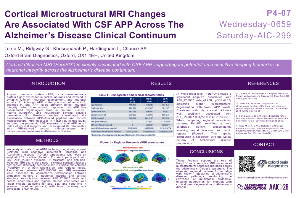
We analyzed data from ADNI including cognitively normal (CN=29), mild cognitive impairment (MCI=43), and Alzheimer’s disease (AD=28) participants who were all amyloid PET positive (Table1). For each participant with CSF APP_P05067 available, T1-structural and diffusion-weighted MRI scans were used to extract cortical thickness and cortical diffusivity perpendicular to cortical minicolumns (PerpPD+ (4) at both whole-brain and regional levels. Associations between APP_P05067 and MRI measures were assessed to characterize relationships between proteomic markers of neuronal integrity and cortical structure. Associations between APP_P05067 levels and cortical whole-brain and regional metrics were tested with linear models, adjusting for age, sex, and variations in scanner model or protocol, with false discovery rate correction (pFDR<0.05).
Results:
At whole-brain level, PerpPD+ showed a significant negative association with APP_P05067 (etap=0.286; pFDR<0.05) indicating higher microstructural degeneration with lower APP levels. Consistent with this, cortical thickness was positively associated with APP_P05067 (etap=0.317; pFDR<0.05).
When comparing regional association patterns, PerpPD+ exhibited a more extensive pattern predominantly involving frontal, temporal, and limbic regions (Figure1). This spatial distribution is consistent with the typical pattern of Alzheimer’s disease progression.
Conclusion:
These findings support the role of PerpPD+ as a sensitive MRI measure of microstructural neurodegeneration across the Alzheimer’s Disease spectrum. The observed regional patterns further align with known trajectories of Alzheimer’s disease pathology, reinforcing the relevance of multimodal proteomic-imaging approaches for characterizing cortical neurodegeneration in Alzheimer’s disease.
References:
1- Orobets KS, Karamyshev AL. Amyloid Precursor Protein and Alzheimer's Disease. Int J Mol Sci. 2023 Sep 30;24(19):14794.
2- Dawkins E, Small DH. Insights into the physiological function of the β-amyloid precursor protein: beyond Alzheimer's disease. J Neurochem. 2014 Jun;129(5):756-69
3- Illán-Gala I, et al. APP-derived peptides reflect neurodegeneration in frontotemporal dementia. Ann Clin TranslNeurol. 2019 Dec;6(12):2518-2530.
4- Torso M,et. A. In Vivo Detection of Changes Related to Cortical Columnar Organization and Neuroinflammation Across the AD Continuum. J Prev Alzheimers Dis. 2022;9(4):769-779.
Comments
Email is required but is not shown publicly.
Loading comments...