4,068 posters, 219 videos, 101 topics, 69 sessions, 14,365 authors, 6,128 institutions
ePostersLive by SciGen Technologies S.A. All rights reserved.
Alzheimer's Association International Conference 2026
July 12-15, 2026 | London, United Kingdom, and Online

1601
Deep Learning-based Cerebrospinal Fluid Segmentation For Normal-pressure Hydrocephalus And Alzheimer’s Disease
Poster Presenter
Authors
Affiliations
Department of Neurology, School of Medicine, Kyungpook National University, Daegu, South Korea, Department of Biomedical Engineering, Daegu Catholic University, Gyeongsan-si, South Korea, Department of Medical Informatics, School of Medicine, Kyungpook National University, Daegu, South Korea
Part of Topic
INTRODUCTION:
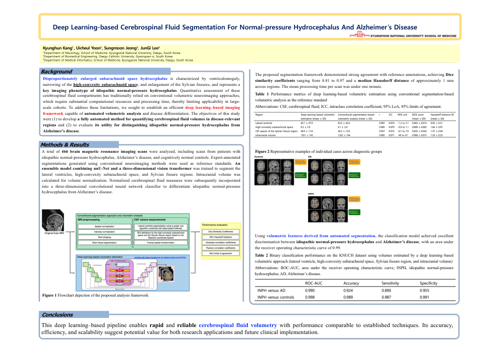
Disproportionately enlarged subarachnoid space hydrocephalus is characterized by ventriculomegaly, narrowing of the high-convexity subarachnoid space, and enlargement of the Sylvian fissures, and represents a key imaging phenotype of idiopathic normal-pressure hydrocephalus. Quantitative assessment of these cerebrospinal fluid compartments has traditionally relied on conventional volumetric neuroimaging approaches, which require substantial computational resources and processing time, thereby limiting applicability in large-scale cohorts. To address these limitations, we sought to establish an efficient deep learning–based imaging framework capable of automated volumetric analysis and disease differentiation. The objectives of this study were (1) to develop a fully automated method for quantifying cerebrospinal fluid volumes in disease-relevant regions and (2) to evaluate its utility for distinguishing idiopathic normal-pressure hydrocephalus from Alzheimer’s disease.
METHODS:
A total of 466 brain magnetic resonance imaging scans were analyzed, including scans from patients with idiopathic normal-pressure hydrocephalus, Alzheimer’s disease, and cognitively normal controls. Expert-annotated segmentations generated using conventional neuroimaging methods were used as reference standards. An ensemble model combining nnU-Net and a three-dimensional vision transformer was trained to segment the lateral ventricles, high-convexity subarachnoid space, and Sylvian fissure regions. Intracranial volume was calculated for volume normalization. Normalized cerebrospinal fluid measures were subsequently incorporated into a three-dimensional convolutional neural network classifier to differentiate idiopathic normal-pressure hydrocephalus from Alzheimer’s disease.
RESULTS:
The proposed segmentation framework demonstrated strong agreement with reference annotations, achieving Dice similarity coefficients ranging from 0.81 to 0.96 and a median Hausdorff distance of approximately 1 mm across regions. The mean processing time per scan was under one minute. Using volumetric features derived from automated segmentation, the classification model achieved excellent discrimination between idiopathic normal-pressure hydrocephalus and Alzheimer’s disease, with an area under the receiver operating characteristic curve of 0.99.
CONCLUSIONS:
This deep learning–based pipeline enables rapid and reliable cerebrospinal fluid volumetry with performance comparable to established techniques. Its accuracy, efficiency, and scalability suggest potential value for both research applications and future clinical implementation.
Comments
Email is required but is not shown publicly.
Loading comments...